Anti-Dystrophin antibody [1808]
| Name | Anti-Dystrophin antibody [1808] |
|---|---|
| Supplier | Abcam |
| Catalog | ab3149 |
| Prices | $400.00 |
| Sizes | 500 µl |
| Host | Mouse |
| Clonality | Monoclonal |
| Isotype | IgG1 |
| Clone | 1808 |
| Applications | Electron microscopy ICC/IF ICC/IF IHC-P WB IHC-F |
| Species Reactivities | Mouse, Rat, Chicken, Human, Xenopus, Torpedo |
| Antigen | Acetylcholine receptor (AChR) enriched membranes and peripheral membrane proteins from Torpedo nobiliana electric organ |
| Description | Mouse Monoclonal |
| Gene | DMD |
| Conjugate | Unconjugated |
| Supplier Page | Shop |
Product images
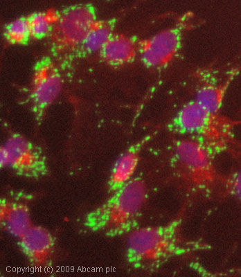 ICC/IF image of ab56078 stained HepG2 cells. The cells were 4% formaldehyde fixed (10 min) and then incubated in 1%BSA / 10% normal goat serum / 0.3M glycine in 0.1% PBS-Tween for 1h to permeabilise the cells and block non-specific protein-protein interactions. The cells were then incubated with the antibody (ab56078, 1µg/ml) overnight at +4°C. The secondary antibody (green) was Alexa Fluor® 488 goat anti-mouse IgG (H+L) used at a 1/1000 dilution for 1h. Alexa Fluor® 594 WGA was used to label plasma membranes (red) at a 1/200 dilution for 1h. DAPI was used to stain the cell nuclei (blue) at a concentration of 1.43µM.
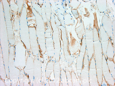 IHC image of Dystrophin staining in Human normal skeletal muscle formalin fixed paraffin embedded tissue section, performed on a Leica BondTM system using the standard protocol F. The section was pre-treated using heat mediated antigen retrieval with sodium citrate buffer (pH6, epitope retrieval solution 1) for 20 mins. The section was then incubated with ab3149, 5µg/ml, for 15 mins at room temperature and detected using an HRP conjugated compact polymer system. DAB was used as the chromogen. The section was then counterstained with haematoxylin and mounted with DPX.For other IHC staining systems (automated and non-automated) customers should optimize variable parameters such as antigen retrieval conditions, primary antibody concentration and antibody incubation times.
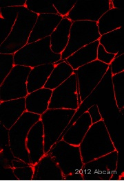 Immunohistochemical analysis of murine skeletal muscle tissue, staining Dystrophin with ab3149.Tissue was fixed with paraformaldehyde, permeabilized with Triton X-100 and blocked with 20 µg/ml BSA for 30 minutes at 25°C. Samples were incubated with primary antibody (1/200 in diluent) for 4 hours at 25°C. An AlexaFluor®546-conjugated anti-mouse monoclonal IgG (1/400) was used as the secondary antibody. See Abreview
Product References
Oxytocin is an age-specific circulating hormone that is necessary for muscle - Oxytocin is an age-specific circulating hormone that is necessary for muscle
Elabd C, Cousin W, Upadhyayula P, Chen RY, Chooljian MS, Li J, Kung S, Jiang KP, Conboy IM. Nat Commun. 2014 Jun 10;5:4082.
Transgenic expression of inclusion body myopathy associated mutant p97/VCP causes - Transgenic expression of inclusion body myopathy associated mutant p97/VCP causes
Weihl CC, Miller SE, Hanson PI, Pestronk A. Hum Mol Genet. 2007 Apr 15;16(8):919-28. Epub 2007 Feb 28.
Localization of dystrophin relative to acetylcholine receptor domains in electric - Localization of dystrophin relative to acetylcholine receptor domains in electric
Sealock R, Butler MH, Kramarcy NR, Gao KX, Murnane AA, Douville K, Froehner SC. J Cell Biol. 1991 Jun;113(5):1133-44.