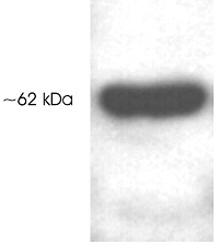
Anti-HDAC1 antibody - C-terminal (ab184651) at 1 µg/ml + HeLa cell extract
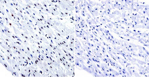
Immunohistochemical analysis of paraffin-embedded Human heart tissue labeling HDAC1 with ab184651 at 1/200 dilution. Tissues were incubated with primary antibody (left) or without primary antibody (negative control, right) overnight at 4°C. Detection was performed using a biotin-conjugated secondary antibody and SA-HRP, followed by colorimetric detection using DAB. Tissues were counterstained with hematoxylin.
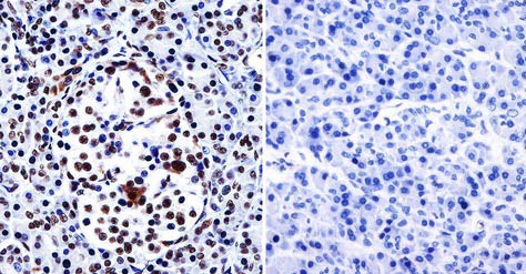
Immunohistochemical analysis of paraffin-embedded Human pancreas tissue labeling HDAC1 with ab184651 at 1/200 dilution. Tissues were incubated with primary antibody (left) or without primary antibody (negative control, right) overnight at 4°C. Detection was performed using a biotin-conjugated secondary antibody and SA-HRP, followed by colorimetric detection using DAB. Tissues were counterstained with hematoxylin.
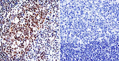
Immunohistochemical analysis of paraffin-embedded Human tonsil tissue labeling HDAC1 with ab184651 at 1/200 dilution. Tissues were incubated with primary antibody (left) or without primary antibody (negative control, right) overnight at 4°C. Detection was performed using a biotin-conjugated secondary antibody and SA-HRP, followed by colorimetric detection using DAB. Tissues were counterstained with hematoxylin.
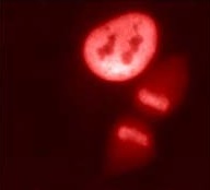
Immunofluorescent analysis of HeLa cells labeling HDAC1 with ab184651 at 2 µg/ml.
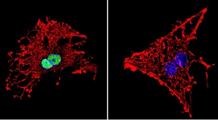
Immunofluroescent analysis of formaldehyde-fixed C6 cells labeling HDAC1 with ab184651 at 1/20 dilution (green). Cells were incubated with primary antibody (left) or without (negative control, right) overnight at 4°C and incubated with a DyLight®488 conjugated secondary antibody. Nuclei were stained with DAPI (blue) and F-Actin with Phalloidin (red).
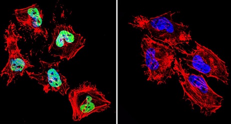
Immunofluroescent analysis of formaldehyde-fixed HeLa cells labeling HDAC1 with ab184651 at 1/20 dilution (green). Cells were incubated with primary antibody (left) or without (negative control, right) overnight at 4°C and incubated with a DyLight®488 conjugated secondary antibody. Nuclei were stained with DAPI (blue) and F-Actin with Phalloidin (red).
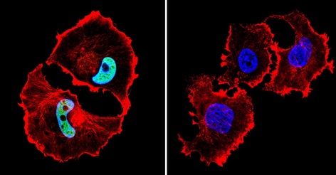
Immunofluorescent analysis of formaldehyde-fixed MCF7 cells labeling HDAC1 with ab184651 at 1/100 dilution (green). Cells were incubated with primary antibody (left) or without (negative control, right) overnight at 4°C and incubated with a DyLight®488 conjugated secondary antibody. Nuclei were stained with DAPI (blue) and F-Actin with Phalloidin (red).