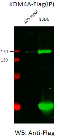
Whole cell lysates derived from 293T transfected with Flag-KDM4A were prepared using PBS+1% Triton plus proteinase inhibitor cocktail and used for IP (1 ug). WB was performed using anti-Flag antibody.
![All lanes : Anti-KDM4A / JHDM3A / JMJD2A antibody [13D6] - ChIP Grade (ab105953) at 1/1000 dilutionLane 1 : As aboveLane 2 : As above](http://www.bioprodhub.com/system/product_images/ab_products/2/sub_3/12210_KDM4A-JHDM3A-JMJD2A-Primary-antibodies-ab105953-2.jpg)
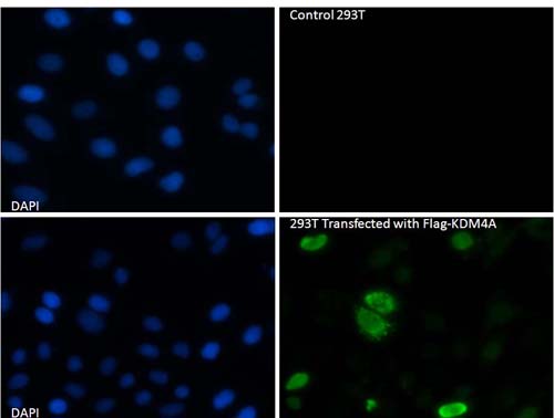
All lanes : Anti-KDM4A / JHDM3A / JMJD2A antibody [13D6] - ChIP Grade (ab105953) at 1/1000 dilutionLane 1 : As aboveLane 2 : As above
ab105935 used at 1/500 dilution
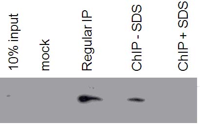
The antibody (1/1000 dilution) was tested for IP of KDM4A in HeLa nuclear extracts under three different conditions: 1) regular (25 mM Tris, pH 7.6, 150 mM KCl, 0.1% NP40, 0.5 mM EDTA, 1 mM DTT; 2) ChIP without SDS (ChIP solution C with 1% Triton and 0.1% deoxycholine); 3) ChIP buffer (as 2 plus 0.1% SDS). 10% input is seen upon longer exposure. SDS presence in this case inhibits IP.
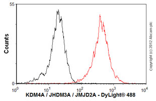
Overlay histogram showing HeLa cells stained with ab105953 (red line). The cells were fixed with 80% methanol (5 min) and then permeabilized with 0.1% PBS-Tween for 20 min. The cells were then incubated in 1x PBS / 10% normal goat serum / 0.3M glycine to block non-specific protein-protein interactions followed by the antibody (ab105953, 0.5µg/1x106 cells) for 30 min at 22ºC. The secondary antibody used was DyLight® 488 goat anti-mouse IgG (H+L) (ab96879) at 1/500 dilution for 30 min at 22ºC. Isotype control antibody (black line) was mouse IgG (1µg/1x106 cells) used under the same conditions. Acquisition of >5,000 events was performed.