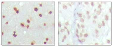
Immunohistochemical staining of paraffin-embedded Human cerebra tissue (left hand image) and human lung carcinoma tissue (right hand image) using ab76362 at 1/200 dilution with DAB staining.
![Anti-MDMX antibody [2D10F4] (ab76362) at 1/500 dilution + Truncated MDMX-His recombinant protein](http://www.bioprodhub.com/system/product_images/ab_products/2/sub_3/20908_mdwb.jpg)
Anti-MDMX antibody [2D10F4] (ab76362) at 1/500 dilution + Truncated MDMX-His recombinant protein
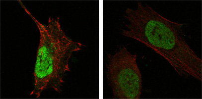
ab76362 at 1/2000 staining MDMX in human Hela (left) and L-02 (right) cells by Immunocytochemistry/ immunofluorescence. An Alexa Fluor® 488 conjugated Goat polyclonal to mouse IgG1 was used as secondary antibody. Green staining in image show positive staining with ab76362, whilst actin filaments were stained red with DY554 phalloidin.
![Overlay histogram showing HeLa cells stained with ab76362 (red line). The cells were fixed with 80% methanol (5 min) and then permeabilized with 0.1% PBS-Tween for 20 min. The cells were then incubated in 1x PBS / 10% normal goat serum / 0.3M glycine to block non-specific protein-protein interactions followed by the antibody (ab76362, 1/100 dilution) for 30 min at 22ºC. The secondary antibody used was DyLight® 488 goat anti-mouse IgG (H+L) (ab96879) at 1/500 dilution for 30 min at 22ºC. Isotype control antibody (black line) was mouse IgG1 [ICIGG1] (ab91353, 2µg/1x106 cells) used under the same conditions. Unlabelled sample (blue line). Acquisition of >5,000 events were collected using a 20mW Argon ion laser (488nm) and 525/30 bandpass filter. This antibody gave a positive signal in HeLa cells fixed with 4% paraformaldehyde (10 min)/permeabilized with 0.1% PBS-Tween for 20 min used under the same conditions.](http://www.bioprodhub.com/system/product_images/ab_products/2/sub_3/20910_MDMX-Primary-antibodies-ab76362-2.jpg)
Overlay histogram showing HeLa cells stained with ab76362 (red line). The cells were fixed with 80% methanol (5 min) and then permeabilized with 0.1% PBS-Tween for 20 min. The cells were then incubated in 1x PBS / 10% normal goat serum / 0.3M glycine to block non-specific protein-protein interactions followed by the antibody (ab76362, 1/100 dilution) for 30 min at 22ºC. The secondary antibody used was DyLight® 488 goat anti-mouse IgG (H+L) (ab96879) at 1/500 dilution for 30 min at 22ºC. Isotype control antibody (black line) was mouse IgG1 [ICIGG1] (ab91353, 2µg/1x106 cells) used under the same conditions. Unlabelled sample (blue line). Acquisition of >5,000 events were collected using a 20mW Argon ion laser (488nm) and 525/30 bandpass filter. This antibody gave a positive signal in HeLa cells fixed with 4% paraformaldehyde (10 min)/permeabilized with 0.1% PBS-Tween for 20 min used under the same conditions.