![Overlay histogram showing A431 cells stained with ab89594 (red line). The cells were fixed with 80% methanol (5 min) and then permeabilized with 0.1% PBS-Tween for 20 min. The cells were then incubated in 1x PBS / 10% normal goat serum / 0.3M glycine to block non-specific protein-protein interactions followed by the antibody (ab89594, 0.1μg/1x106 cells) for 30 min at 22°C. The secondary antibody used was Alexa Fluor® 488 goat anti-mouse IgG (H&L) (ab150113) at 1/2000 dilution for 30 min at 22°C. Isotype control antibody (black line) was mouse IgG2b [PLPV219] (ab91366, 1μg/1x106 cells) used under the same conditions. Unlabelled sample (blue line) was also used as a control. Acquisition of >5,000 events were collected using a 20mW Argon ion laser (488nm) and 525/30 bandpass filter.](http://www.bioprodhub.com/system/product_images/ab_products/2/sub_3/26966_ab89594-4-ab89594FC.jpg)
Overlay histogram showing A431 cells stained with ab89594 (red line). The cells were fixed with 80% methanol (5 min) and then permeabilized with 0.1% PBS-Tween for 20 min. The cells were then incubated in 1x PBS / 10% normal goat serum / 0.3M glycine to block non-specific protein-protein interactions followed by the antibody (ab89594, 0.1μg/1x106 cells) for 30 min at 22°C. The secondary antibody used was Alexa Fluor® 488 goat anti-mouse IgG (H&L) (ab150113) at 1/2000 dilution for 30 min at 22°C. Isotype control antibody (black line) was mouse IgG2b [PLPV219] (ab91366, 1μg/1x106 cells) used under the same conditions. Unlabelled sample (blue line) was also used as a control. Acquisition of >5,000 events were collected using a 20mW Argon ion laser (488nm) and 525/30 bandpass filter.
![Anti-Myosin Light Chain 2 antibody [AT3B2] (ab89594) at 1/1000 dilution + rat heart extracts at 60 µgSecondarygoat anti-mouse HRPdeveloped using the ECL technique](http://www.bioprodhub.com/system/product_images/ab_products/2/sub_3/26967_Myosin-Light-Chain-2-Primary-antibodies-ab89594-1.jpg)
Anti-Myosin Light Chain 2 antibody [AT3B2] (ab89594) at 1/1000 dilution + rat heart extracts at 60 µgSecondarygoat anti-mouse HRPdeveloped using the ECL technique
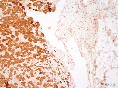
ab98594 staining Myosin Light Chain 2 in Rabbit heart tissue sections by Immunohistochemistry (IHC-P - paraformaldehyde-fixed, paraffin-embedded sections). Tissue was fixed with formaldehyde and blocked with 10% serum for 60 minutes; antigen retrieval was by heat mediation in a Tris/EDTA buffer. Samples were incubated with primary antibody (1/500 in PBS-T) for 1 hour at 37°C. A HRP-conjugated Goat anti-mouse polyclonal (1/1000) was used as the secondary antibody.See Abreview
![Anti-Myosin Light Chain 2 antibody [AT3B2] (ab89594) at 1/2000 dilution + Rabbit heart tissue lysate at 10 µgSecondaryHRP conjugated goat anti-mouse at 1/5000 dilutiondeveloped using the ECL techniquePerformed under reducing conditions.](http://www.bioprodhub.com/system/product_images/ab_products/2/sub_3/26969_Myosin-Light-Chain-2-Primary-antibodies-ab89594-4.jpg)
Anti-Myosin Light Chain 2 antibody [AT3B2] (ab89594) at 1/2000 dilution + Rabbit heart tissue lysate at 10 µgSecondaryHRP conjugated goat anti-mouse at 1/5000 dilutiondeveloped using the ECL techniquePerformed under reducing conditions.