![All lanes : Anti-NDUFAF4 [EPR15931] antibody (ab191414) at 1/10000 dilutionLane 1 : T47D cell lysateLane 2 : Raji cell lysateLane 3 : K562 cell lysateLane 4 : MDA-MB-435 cell lysateLysates/proteins at 20 µg per lane.SecondaryGoat Anti-Rabbit IgG, (H+L), Peroxidase conjugated at 1/1000 dilution](http://www.bioprodhub.com/system/product_images/ab_products/2/sub_3/28397_ab191414-228636-ab191414-wb-1.jpg)
All lanes : Anti-NDUFAF4 [EPR15931] antibody (ab191414) at 1/10000 dilutionLane 1 : T47D cell lysateLane 2 : Raji cell lysateLane 3 : K562 cell lysateLane 4 : MDA-MB-435 cell lysateLysates/proteins at 20 µg per lane.SecondaryGoat Anti-Rabbit IgG, (H+L), Peroxidase conjugated at 1/1000 dilution
![Anti-NDUFAF4 [EPR15931] antibody (ab191414) at 1/1000 dilution + MCF7 cell lysate at 10 µgSecondaryGoat Anti-Rabbit IgG, (H+L), Peroxidase conjugated at 1/1000 dilution](http://www.bioprodhub.com/system/product_images/ab_products/2/sub_3/28398_ab191414-228637-ab191414-wb-2.jpg)
Anti-NDUFAF4 [EPR15931] antibody (ab191414) at 1/1000 dilution + MCF7 cell lysate at 10 µgSecondaryGoat Anti-Rabbit IgG, (H+L), Peroxidase conjugated at 1/1000 dilution
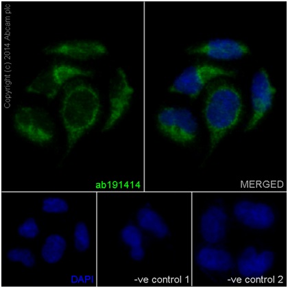
Immunofluorescence analysis of HeLa cells labeling NDUFAF4 using ab191414 at 1/150 dilution (9.5 μg/mL). A Goat anti Rabbit IgG (Alexa Fluor488) at 1/200 dilution (ab150077) was used as secondary antibody. Cells were fixed with 4% Paraformaldehyde and permealized with 0.1% triton X-100. Counterstain: DAPI. The 2 negative cotrols images were obtained using Goat anti Mouse IgG (Alexa Fluor®594) at 1:400 dilution as secondary antibody.
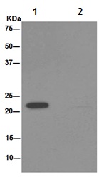
Immunoprecipitation analysis of MDA-MB-435 cell lysate labeling NDUFAF4 using ab191414 at 1/90 dilution (Lane 1). An Anti-Rabbit IgG (HRP), specific to the non-reduced form of IgG at 1/1500 was used as secondary antibody. Lane 2: PBS instead of MDA-MB-435 cell lysate.