![All lanes : Anti-Nogo antibody [EPR12265] (ab180847) at 1/1000 dilutionLane 1 : U87-MG cell lysateLane 2 : SH-SY5Y cell lysateLane 3 : HeLa cell lysateLysates/proteins at 10 µg per lane.SecondaryGoat anti-rabbit HRP at 1/2000 dilutiondeveloped using the ECL technique](http://www.bioprodhub.com/system/product_images/ab_products/2/sub_4/1233_ab180847-210296-ab180847WB.jpg)
All lanes : Anti-Nogo antibody [EPR12265] (ab180847) at 1/1000 dilutionLane 1 : U87-MG cell lysateLane 2 : SH-SY5Y cell lysateLane 3 : HeLa cell lysateLysates/proteins at 10 µg per lane.SecondaryGoat anti-rabbit HRP at 1/2000 dilutiondeveloped using the ECL technique

Immunohistochemical analysis of formalin-fixed, paraffin-embedded, Human brain tissue labeling Nogo with ab180847 at a 1/50 dilution.
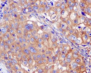
Immunohistochemical analysis of formalin-fixed, paraffin-embedded, Human breast carcinoma tissue labeling Nogo with ab180847 at a 1/50 dilution.
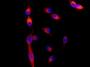
Immunofluorescence analysis of U87-MG cells labeling Nogo with ab180847 at a 1/100 dilution.
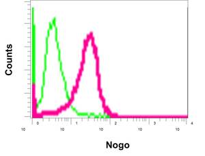
Flow cytometry analysis of permeabilized HeLa cells labeling Nogo (red) with ab180847 at a 1/10 dilution, or negative control rabbit IgG (green)
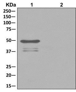
Western blot analysis on Immunoprecipitation pellet from either 1) HeLa cell lysate, or 2) 1xPBS (negative control); showing Nogo, using ab180847 at 1/10 dilution and HRP-conjugated anti-rabbit IgG preferentially detecting the non-reduced form of rabbit IgG.