![All lanes : Anti-Nucleobindin 2 antibody [EPR13159] (ab181159) at 1/1000 dilutionLane 1 : MCF-7 cell lysateLane 2 : HepG2 cell lysateLane 3 : U87-MG cell lysateLysates/proteins at 10 µg per lane.SecondaryGoat Anti-Rabbit IgG, (H+L), Peroxidase conjugate at 1/1000 dilution](http://www.bioprodhub.com/system/product_images/ab_products/2/sub_4/2553_ab181159-214676-ab181159WB.jpg)
All lanes : Anti-Nucleobindin 2 antibody [EPR13159] (ab181159) at 1/1000 dilutionLane 1 : MCF-7 cell lysateLane 2 : HepG2 cell lysateLane 3 : U87-MG cell lysateLysates/proteins at 10 µg per lane.SecondaryGoat Anti-Rabbit IgG, (H+L), Peroxidase conjugate at 1/1000 dilution
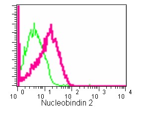
Flow cytometric analysis of 2% paraformaldehyde-fixed HepG2 cells labeling Nucleobindin 2 with ab181159 at 1/500 dilution (red) compared to a Rabbit monoclonal IgG isotype control (green), followed by Goat anti-rabbit IgG (FITC) secondary antibody at 1/150 dilution.
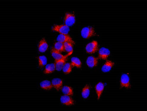
Immunofluorescent analysis of 4% paraformaldehyde-fixed MCF-7 cells labeling Nucleobindin 2 with ab181159 at 1/250 dilution followed by Goat anti rabbit IgG (Alexa Fluor® 555) secondary antibody at 1/200 dilution. Counter stained with DAPI.
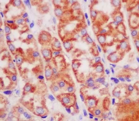
Immunohistochemical analysis of paraffin-embedded Human stomach tissue labeling Nucleobindin 2 with ab181159 at 1/250 dilution followed by prediluted HRP Polymer for Rabbit IgG. Counter stained with Hematoxylin.
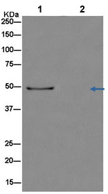
Western blot analysis of Nucleobindin 2 in Human stomach lysate immunoprecipitated with ab181159 at 1/50 dilution (Lane 1). Lane 2: Negative control.Secondary: Anti-Rabbit IgG (HRP), specific to the non-reduced form of IgG at 1/1500 dilution.