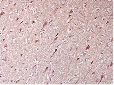
IHC-P image of PERK staining on human cortical sections using ab79483 (1:500). The tissue was fixed in formaldehyde and the sections were then subjected to heat mediated antigen retrieval using citric acid and permeabilized using Triton-X. The sections were them blocked using 2% BSA for 1 hour at 20°C. ab79483 was diluted 1:500 and incubated with the sections for 18 hours at 20°C. The secondary antibody used was HRP conjugated Goat polyclonal to Rabbit IgG (1:500)See Abreview
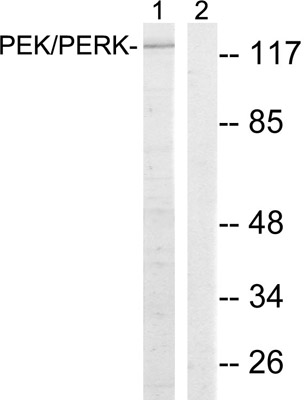
All lanes : Anti-PERK antibody (ab79483) at 1/500 dilutionLane 1 : extracts from MCF-7 cellsLane 2 : extracts from MCF-7 cells with immunising peptide at 10 µgLysates/proteins at 30 µg per lane.
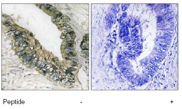
Immunohistochemistry analysis of paraffin-embedded human colon carcinoma tissue, stained with ab79483 at 1/50-1/100 dilution, in the absence and presence of the immunising peptide