Anti-RhoA antibody
| Name | Anti-RhoA antibody |
|---|---|
| Supplier | Abcam |
| Catalog | ab86297 |
| Prices | $378.00 |
| Sizes | 100 µg |
| Host | Rabbit |
| Clonality | Polyclonal |
| Isotype | IgG |
| Applications | ICC/IF ICC/IF WB |
| Species Reactivities | Mouse, Rat, Human, Chicken, Bovine, Dog, Zebrafish, Orangutan |
| Antigen | Synthetic peptide conjugated to KLH derived from within residues 150 to the C-terminus of Human RhoA |
| Description | Rabbit Polyclonal |
| Gene | RHOA |
| Conjugate | Unconjugated |
| Supplier Page | Shop |
Product images
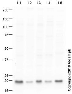 All lanes : Anti-RhoA antibody (ab86297) at 1 µg/mlLane 1 : HL60 (Human promyelocytic leukemia cell line) Whole Cell Lysate Lane 2 : MCF7 (Human breast adenocarcinoma cell line) Whole Cell LysateLane 3 : MDA-MB-231 (Human breast adenocarcinoma cell line) Whole Cell Lysate Lane 4 : HeLa (Human epithelial carcinoma cell line) Whole Cell LysateLane 5 : Human Platelet (Human adult normal cell line) Whole Cell LysateLysates/proteins at 10 µg per lane.SecondaryGoat Anti-Rabbit IgG H&L (HRP) preadsorbed (ab97080) at 1/5000 dilutiondeveloped using the ECL techniquePerformed under reducing conditions.
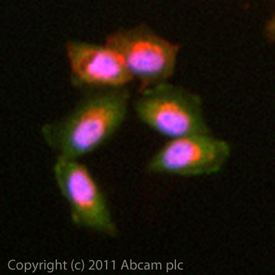 ICC/IF image of ab86297 stained MCF7 cells. The cells were 4% PFA fixed (10 min) and then incubated in 1%BSA / 10% normal goat serum / 0.3M glycine in 0.1% PBS-Tween for 1h to permeabilise the cells and block non-specific protein-protein interactions. The cells were then incubated with the antibody (ab86297, 10µg/ml) overnight at +4°C. The secondary antibody (green) was ab96899 Dylight 488 goat anti-rabbit IgG (H+L) used at a 1/250 dilution for 1h. Alexa Fluor® 594 WGA was used to label plasma membranes (red) at a 1/200 dilution for 1h. DAPI was used to stain the cell nuclei (blue) at a concentration of 1.43µM.
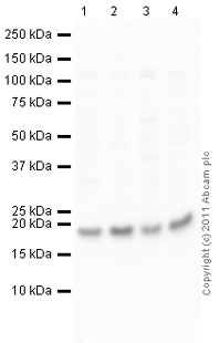 All lanes : Anti-RhoA antibody (ab86297) at 1 µg/mlLane 1 : Kidney (Mouse) Tissue LysateLane 2 : Lung (Mouse) Whole Cell Lysate - normal tissue (ab29297)Lane 3 : Kidney (Rat) Tissue LysateLane 4 : Lung (Rat) Tissue Lysate Lysates/proteins at 10 µg per lane.SecondaryGoat Anti-Rabbit IgG H&L (HRP) preadsorbed (ab97080) at 1/5000 dilutiondeveloped using the ECL techniquePerformed under reducing conditions.
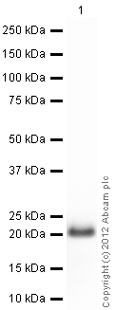 Anti-RhoA antibody (ab86297) at 1 µg/ml + Human RhoA full length protein (ab101594) at 0.01 µgSecondaryGoat Anti-Rabbit IgG H&L (HRP) preadsorbed (ab97080) at 1/5000 dilutiondeveloped using the ECL techniquePerformed under reducing conditions.Exposure time : 10 seconds
Product References
Coronaviruses induce entry-independent, continuous macropinocytosis. - Coronaviruses induce entry-independent, continuous macropinocytosis.
Freeman MC, Peek CT, Becker MM, Smith EC, Denison MR. MBio. 2014 Aug 5;5(4):e01340-14.
RhoA localization with caveolin-1 regulates vascular contractions to serotonin. - RhoA localization with caveolin-1 regulates vascular contractions to serotonin.
Nuno DW, England SK, Lamping KG. Am J Physiol Regul Integr Comp Physiol. 2012 Nov 1;303(9):R959-67. doi: