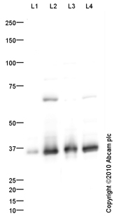
All lanes : Anti-RPL5 antibody (ab86863) at 1 µg/mlLane 1 : Liver (Human)Tissue Lysate - Fetal tissueLane 2 : HepG2 (Human hepatocellular liver carcinoma cell line) Whole Cell LysateLane 3 : MEL-1 (Human embryonic stem cell, male cell line) Whole Cell Lysate (ab27198)Lane 4 : Jurkat (Human T cell lymphoblast-like cell line) Whole Cell LysateLysates/proteins at 10 µg per lane.SecondaryGoat polyclonal to Rabbit IgG - H&L - Pre-Adsorbed (HRP) at 1/3000 dilutiondeveloped using the ECL techniquePerformed under reducing conditions.
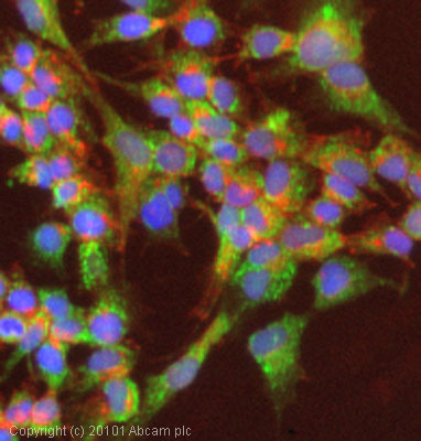
ICC/IF image of ab86863 stained HepG2 cells. The cells were 4% PFA fixed (10 min) and then incubated in 1%BSA / 10% normal goat serum / 0.3M glycine in 0.1% PBS-Tween for 1h to permeabilise the cells and block non-specific protein-protein interactions. The cells were then incubated with the antibody (ab86863, 5µg/ml) overnight at +4°C. The secondary antibody (green) was Alexa Fluor® 488 goat anti-rabbit IgG (H+L) used at a 1/1000 dilution for 1h. Alexa Fluor® 594 WGA was used to label plasma membranes (red) at a 1/200 dilution for 1h. DAPI was used to stain the cell nuclei (blue) at a concentration of 1.43µM. This antibody also gave a positive result in 4% PFA fixed (10 min) HeLa cells at 5µg/ml, and in 100% methanol fixed (5 min) HeLa, HepG2 and MCF7 cells at 5µg/ml
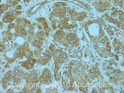
IHC image of RPL5 staining in human liver carcinoma formalin fixed paraffin embedded tissue section, performed on a Leica BondTM system using the standard protocol F. The section was pre-treated using heat mediated antigen retrieval with sodium citrate buffer (pH6, epitope retrieval solution 1) for 20 mins. The section was then incubated with ab86863, 1µg/ml, for 15 mins at room temperature and detected using an HRP conjugated compact polymer system. DAB was used as the chromogen. The section was then counterstained with haematoxylin and mounted with DPX.
![RPL5 was immunoprecipitated using 0.5mg HepG2 whole cell extract, 5µg of Rabbit polyclonal to RPL5 and 50µl of protein G magnetic beads (+). No antibody was added to the control (-). The antibody was incubated under agitation with Protein G beads for 10min, HepG2 whole cell extract lysate diluted in RIPA buffer was added to each sample and incubated for a further 10min under agitation.Proteins were eluted by addition of 40µl SDS loading buffer and incubated for 10min at 70oC; 10µl of each sample was separated on a SDS PAGE gel, transferred to a nitrocellulose membrane, blocked with 5% BSA and probed with ab86863.Secondary: Mouse monoclonal [SB62a] Secondary Antibody to Rabbit IgG light chain (HRP) (ab99697).Band: 34kDa, non specific band - 68kDa as present in test (lane 1) and control (lane 2); 68kDa: We are unsure as to the identity of this extra band; RPL5](http://www.bioprodhub.com/system/product_images/ab_products/2/sub_4/24854_RPL5-Primary-antibodies-ab86863-5.jpg)
RPL5 was immunoprecipitated using 0.5mg HepG2 whole cell extract, 5µg of Rabbit polyclonal to RPL5 and 50µl of protein G magnetic beads (+). No antibody was added to the control (-). The antibody was incubated under agitation with Protein G beads for 10min, HepG2 whole cell extract lysate diluted in RIPA buffer was added to each sample and incubated for a further 10min under agitation.Proteins were eluted by addition of 40µl SDS loading buffer and incubated for 10min at 70oC; 10µl of each sample was separated on a SDS PAGE gel, transferred to a nitrocellulose membrane, blocked with 5% BSA and probed with ab86863.Secondary: Mouse monoclonal [SB62a] Secondary Antibody to Rabbit IgG light chain (HRP) (ab99697).Band: 34kDa, non specific band - 68kDa as present in test (lane 1) and control (lane 2); 68kDa: We are unsure as to the identity of this extra band; RPL5