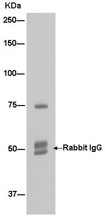
Western blot analysis of HepG2 cell lysate immunoprecipitated using ab181856 at 1/50 dilution. Goat Anti-Rabbit IgG, (H+L), Peroxidase conjugate secondary antibody was used at 1/1000 dilution.
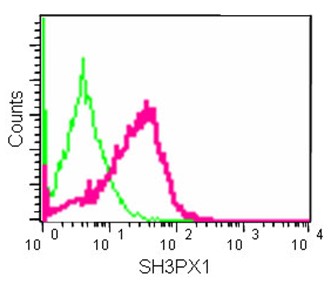
Flow cytometric analysis of 2% paraformaldehyde-fixed HeLa cells labeling SH3PX1 with ab181856 at 1/10 dilution (red) compared to a Rabbit IgG monoclonal isotype control (green), followed by Goat anti rabbit IgG (FITC) secondary antibody at 1/150 dilution.
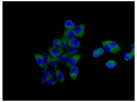
Immunofluorescent analysis of 4% paraformaldehyde-fixed HCT-116 cells labeling SH3PX1 with ab181856 at 1/100 dilution, followed by Goat anti rabbit IgG (Alexa Fluor®488) secondary antibody at 1/200 dilution. Counter stained with Dapi.
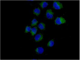
Immunofluorescent analysis of acetone-fixed HeLa cells labeling SH3PX1 with ab181856 at 1/100 dilution, followed by Goat anti rabbit IgG (Alexa Fluor®488) secondary antibody at 1/200 dilution. Counter stained with Dapi.

Immunohistochemical analysis of paraffin-embedded Human colon tissue labeling SH3PX1 with ab181856 at 1/250 dilution, followed by prediluted HRP Polymer for Rabbit IgG. Counter stained with Hematoxylin.
![Anti-SH3PX1 antibody [EPR14399] (ab181856) at 1/10000 dilution + HCT-116 cell lysate at 20 µgSecondaryGoat Anti-Rabbit IgG, (H+L), Peroxidase conjugate at 1/1000 dilution](http://www.bioprodhub.com/system/product_images/ab_products/2/sub_4/29582_ab181856-213165-ab1818562.jpg)
Anti-SH3PX1 antibody [EPR14399] (ab181856) at 1/10000 dilution + HCT-116 cell lysate at 20 µgSecondaryGoat Anti-Rabbit IgG, (H+L), Peroxidase conjugate at 1/1000 dilution
![Anti-SH3PX1 antibody [EPR14399] (ab181856) at 1/50000 dilution + HeLa cell lysate at 20 µgSecondaryGoat Anti-Rabbit IgG, (H+L), Peroxidase conjugate at 1/1000 dilution](http://www.bioprodhub.com/system/product_images/ab_products/2/sub_4/29583_ab181856-213164-ab181856.jpg)
Anti-SH3PX1 antibody [EPR14399] (ab181856) at 1/50000 dilution + HeLa cell lysate at 20 µgSecondaryGoat Anti-Rabbit IgG, (H+L), Peroxidase conjugate at 1/1000 dilution