![All lanes : Anti-SLC40A1 antibody [mAbcam93438] (ab93438) at 1/1000 dilutionLane 1 : 1 month old mouse liverLane 2 : 3 month old mouse liverLane 3 : 3 month old mouse Fe+ liverLane 4 : 6 month old mouse Fe+ liverLysates/proteins at 20 µg per lane.SecondaryGoat Polyclonal to mouse IgG conjugated to HRPdeveloped using the ECL techniquePerformed under reducing conditions.](http://www.bioprodhub.com/system/product_images/ab_products/2/sub_5/1255_SLC40A1-Primary-antibodies-ab93438-3.jpg)
All lanes : Anti-SLC40A1 antibody [mAbcam93438] (ab93438) at 1/1000 dilutionLane 1 : 1 month old mouse liverLane 2 : 3 month old mouse liverLane 3 : 3 month old mouse Fe+ liverLane 4 : 6 month old mouse Fe+ liverLysates/proteins at 20 µg per lane.SecondaryGoat Polyclonal to mouse IgG conjugated to HRPdeveloped using the ECL techniquePerformed under reducing conditions.
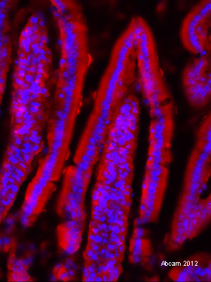
IHC-Fr image of SLC40A1 staining on mouse duodenum using ab93438 (1:1000). The sections were blocked using donkey serum. The sections were incubated with ab93438 at 1:1000 dilution. The secondary antibody used was Donkey polyclonal to mouse IgG conjugated to Alexa Fluor 594 (1:1000).
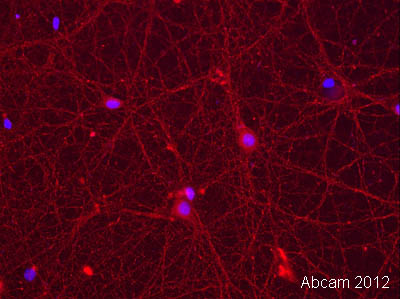
ICC/IF image of SLC40A1 staining in mouse hippocampal culture using ab93438 (1:1000). The cells were permeabilized using 0.1% TritonX in 0.1% PBS. The cells were then incubated with 10% donkey serum for 1 hour at 24°C. ab93438 (1:1000) was incubated for 4 hours at 24°C. the secondary antibody used was donkey polyclonal to Mouse IgG conjugated to Alexa Fluor 568.
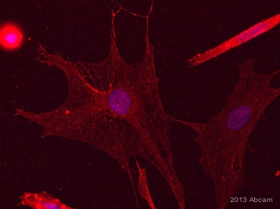
ICC/IF image of SLC40A1 staining in Rat astrocyte culture using ab93438 (1:1000). The cells were permeabilized using 0.1% TritonX in 0.1% PBS. The cells were then incubated with 10% donkey serum for 1 hour at 24°C. ab93438 (1:1000) was incubated for 4 hours at 24°C. the secondary antibody used was donkey polyclonal to Rat IgG conjugated to Alexa Fluor 568.See Abreview