Anti-TATA binding protein TBP antibody [mAbcam 51841] - Nuclear Loading Control and ChIP Grade
| Name | Anti-TATA binding protein TBP antibody [mAbcam 51841] - Nuclear Loading Control and ChIP Grade |
|---|---|
| Supplier | Abcam |
| Catalog | ab51841 |
| Prices | $404.00, $809.00 |
| Sizes | 100 µg, 250 µg |
| Host | Mouse |
| Clonality | Monoclonal |
| Isotype | IgG |
| Clone | mAbcam 51841 |
| Applications | WB ChIP ICC/IF ICC/IF FC ChIP ChIP IP IHC-P |
| Species Reactivities | Mouse, Rat, Human, Chicken, Bovine, Xenopus, Chimpanzee, Zebrafish |
| Antigen | Synthetic peptide conjugated to KLH derived from within residues 1 - 100 of Human TATA binding protein TBP |
| Description | Mouse Monoclonal |
| Gene | TBP |
| Conjugate | Unconjugated |
| Supplier Page | Shop |
Product images
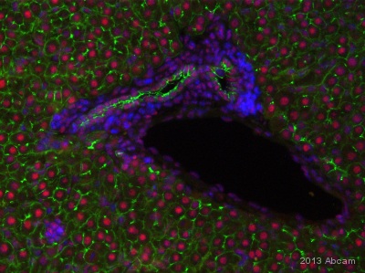 ab51841 staining TATA binding protein TBP and ab15102 staining Claudin3 in Mouse liver tissue sections by Immunohistochemistry (IHC-P - paraformaldehyde-fixed, paraffin-embedded sections). Tissue was fixed with paraformaldehyde and blocked with 10% serum for 30 minutes at 25°C; antigen retrieval was by heat mediation in a citrate buffer pH6. Samples were incubated with primary antibodies ab51841 and ab15102 (1/400 and 1/300 respectively in blocking buffer) for 16 hours at 4°C. A Cy3-conjugated Goat anti-mouse IgG polyclonal (1/200) was used as the secondary antibody. See Abreview
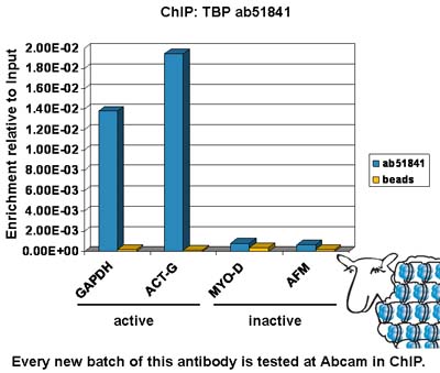 Chromatin was prepared from Hela cells according to the Abcam X-ChIP protocol. Cells were fixed with formaldehyde for 10 min. The ChIP was performed with 25 µg of chromatin, 5 µg of ab51841 (blue), and 20 µl of Protein A/G sepharose beads. No antibody was added to the beads control (yellow). The immunoprecipitated DNA was quantified by real time PCR (Taqman or sybr green approach). Primers and probes are located within 1 kb of the transcription start site.
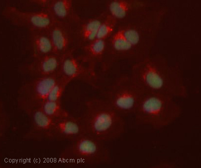 ICC/IF image of ab51841 stained human HEK 293 cells. The cells were 4% PFA fixed (10 min), permabilised in 0.1% PBS-Tween (20 min) and incubated with the antibody (ab51841, 1µg/ml) for 1h at room temperature. 1%BSA / 10% normal goat serum / 0.3M glycine was used to block non-specific protein-protein interactions. The secondary antibody (green) was Alexa Fluor® 488 goat anti-mouse IgG (H+L) used at a 1/1000 dilution for 1h. Alexa Fluor® 594 WGA was used to label plasma membranes (red). DAPI was used to stain the cell nuclei (blue). This antibody also gave a positive IF result in HeLa, HepG2 and MCF7 cells.
![All lanes : Anti-TATA binding protein TBP antibody [mAbcam 51841] - Nuclear Loading Control and ChIP Grade (ab51841) at 5 µg/mlLane 1 : HeLa (Human epithelial carcinoma cell line) Cytoplasmic Lysate at 10 µgLane 2 : HeLa (Human epithelial carcinoma cell line) Nuclear Lysate at 10 µgLane 3 : HeLa (Human epithelial carcinoma cell line) Nuclear Lysate at 20 µgSecondaryRabbit polyclonal to Mouse IgG - H&L (HRP) at 1/3000 dilutionPerformed under reducing conditions.](http://www.bioprodhub.com/system/product_images/ab_products/2/sub_5/8530_ab51841_3.jpg) All lanes : Anti-TATA binding protein TBP antibody [mAbcam 51841] - Nuclear Loading Control and ChIP Grade (ab51841) at 5 µg/mlLane 1 : HeLa (Human epithelial carcinoma cell line) Cytoplasmic Lysate at 10 µgLane 2 : HeLa (Human epithelial carcinoma cell line) Nuclear Lysate at 10 µgLane 3 : HeLa (Human epithelial carcinoma cell line) Nuclear Lysate at 20 µgSecondaryRabbit polyclonal to Mouse IgG - H&L (HRP) at 1/3000 dilutionPerformed under reducing conditions.
All lanes : Anti-TATA binding protein TBP antibody [mAbcam 51841] - Nuclear Loading Control and ChIP Grade (ab51841) at 5 µg/mlLane 1 : HeLa (Human epithelial carcinoma cell line) Cytoplasmic Lysate at 10 µgLane 2 : HeLa (Human epithelial carcinoma cell line) Nuclear Lysate at 10 µgLane 3 : HeLa (Human epithelial carcinoma cell line) Nuclear Lysate at 20 µgSecondaryRabbit polyclonal to Mouse IgG - H&L (HRP) at 1/3000 dilutionPerformed under reducing conditions.
 IHC image of TATA binding protein TBP staining in human breast carcinoma FFPE section, performed on a BondTM system using the standard protocol F. The section was pre-treated using heat mediated antigen retrieval with sodium citrate buffer (pH6, epitope retrieval solution 1) for 20 mins. The section was then incubated with ab51841, 1µg/ml, for 15 mins at room temperature and detected using an HRP conjugated compact polymer system. DAB was used as the chromogen. The section was then counterstained with haematoxylin and mounted with DPX.
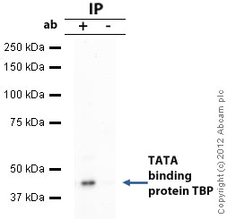 TBP was immunoprecipitated using 0.5mg Hela whole cell extract, 10µg of Mouse monoclonal to TBP and 50µl of protein G magnetic beads (+). No antibody was added to the control (-). The antibody was incubated under agitation with Protein G beads for 10min, Hela whole cell extract lysate diluted in RIPA buffer was added to each sample and incubated for a further 10min under agitation.Proteins were eluted by addition of 40µl SDS loading buffer and incubated for 10min at 70oC; 10µl of each sample was separated on a SDS PAGE gel, transferred to a nitrocellulose membrane, blocked with 5% BSA and probed with ab51841.Secondary: Goat polyclonal to mouse IgG light chain specific (HRP) at 1/20,000 dilution.Band: 40kDa: TATA binding protein TBP.
 Overlay histogram showing HeLa cells stained with ab51841 (red line). The cells were fixed with 80% methanol (5 min) and then permeabilized with 0.1% PBS-Tween for 20 min. The cells were then incubated in 1x PBS / 10% normal goat serum / 0.3M glycine to block non-specific protein-protein interactions followed by the antibody (ab51841, 1µg/1x106 cells) for 30 min at 22ºC. The secondary antibody used was DyLight® 488 goat anti-mouse IgG (H+L) (ab96879) at 1/500 dilution for 30 min at 22ºC. Isotype control antibody (black line) was mouse IgG (1µg/1x106 cells) used under the same conditions. Acquisition of >5,000 events was performed.
Product References
Core promoter factor TAF9B regulates neuronal gene expression. - Core promoter factor TAF9B regulates neuronal gene expression.
Herrera FJ, Yamaguchi T, Roelink H, Tjian R. Elife. 2014 Jul 8;3:e02559.
Reconstitution of human rRNA gene transcription in mouse cells by a complete SL1 - Reconstitution of human rRNA gene transcription in mouse cells by a complete SL1
Murano K, Okuwaki M, Momose F, Kumakura M, Ueshima S, Newbold RF, Nagata K. J Cell Sci. 2014 Aug 1;127(Pt 15):3309-19.
The adaptor CRADD/RAIDD controls activation of endothelial cells by - The adaptor CRADD/RAIDD controls activation of endothelial cells by
Qiao H, Liu Y, Veach RA, Wylezinski L, Hawiger J. J Biol Chem. 2014 Aug 8;289(32):21973-83.
Is nuclear factor erythroid 2-related factor 2 responsible for sex differences in - Is nuclear factor erythroid 2-related factor 2 responsible for sex differences in
Rohrer PR, Rudraiah S, Goedken MJ, Manautou JE. Drug Metab Dispos. 2014 Oct;42(10):1663-74.
Adenovirus L-E1A activates transcription through mediator complex-dependent - Adenovirus L-E1A activates transcription through mediator complex-dependent
Vijayalingam S, Chinnadurai G. J Virol. 2013 Mar;87(6):3425-34.
Circadian clock protein cryptochrome regulates the expression of proinflammatory - Circadian clock protein cryptochrome regulates the expression of proinflammatory
Narasimamurthy R, Hatori M, Nayak SK, Liu F, Panda S, Verma IM. Proc Natl Acad Sci U S A. 2012 Jul 31;109(31):12662-7. doi:
AMPK and substrate availability regulate creatine transport in cultured - AMPK and substrate availability regulate creatine transport in cultured
Darrabie MD, Arciniegas AJ, Mishra R, Bowles DE, Jacobs DO, Santacruz L. Am J Physiol Endocrinol Metab. 2011 May;300(5):E870-6. doi:
Selenoprotein K knockout mice exhibit deficient calcium flux in immune cells and - Selenoprotein K knockout mice exhibit deficient calcium flux in immune cells and
Verma S, Hoffmann FW, Kumar M, Huang Z, Roe K, Nguyen-Wu E, Hashimoto AS, Hoffmann PR. J Immunol. 2011 Feb 15;186(4):2127-37.
Comprehensive study in the inhibitory effect of berberine on gene transcription, - Comprehensive study in the inhibitory effect of berberine on gene transcription,
Wang Y, Kheir MM, Chai Y, Hu J, Xing D, Lei F, Du L. PLoS One. 2011;6(8):e23495.
The phosphatase interactor NIPP1 regulates the occupancy of the histone - The phosphatase interactor NIPP1 regulates the occupancy of the histone
Van Dessel N, Beke L, Gornemann J, Minnebo N, Beullens M, Tanuma N, Shima H, Van Eynde A, Bollen M. Nucleic Acids Res. 2010 Nov;38(21):7500-12.