Anti-TATA binding protein TBP antibody - Nuclear Loading Control and ChIP Grade
| Name | Anti-TATA binding protein TBP antibody - Nuclear Loading Control and ChIP Grade |
|---|---|
| Supplier | Abcam |
| Catalog | ab63766 |
| Prices | $403.00, $806.00 |
| Sizes | 100 µg, 250 µg |
| Host | Rabbit |
| Clonality | Polyclonal |
| Isotype | IgG |
| Applications | ICC/IF ICC/IF IP ChIP IHC-P WB |
| Species Reactivities | Mouse, Rat, Human, Chicken, Bovine |
| Antigen | Synthetic peptide conjugated to KLH derived from within residues 1 - 100 of Human TATA binding protein TBP |
| Description | Rabbit Polyclonal |
| Gene | TBP |
| Conjugate | Unconjugated |
| Supplier Page | Shop |
Product images
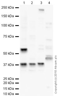 All lanes : Anti-TATA binding protein TBP antibody - Nuclear Loading Control and ChIP Grade (ab63766) at 1 µg/mlLane 1 : Testis (Mouse) Tissue LysateLane 2 : NIH 3T3 (Mouse embryonic fibroblast cell line) Whole Cell LysateLane 3 : Testis (Rat) Tissue LysateLane 4 : HepG2 (Human hepatocellular liver carcinoma cell line) Nuclear Lysate (ab14660)Lysates/proteins at 10 µg per lane.SecondaryGoat polyclonal to Rabbit IgG - H&L - Pre-Adsorbed (HRP) at 1/3000 dilutiondeveloped using the ECL techniquePerformed under reducing conditions.
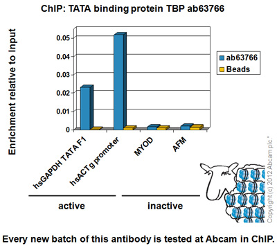 Chromatin was prepared from HeLa cells according to the Abcam X-ChIP protocol. Cells were fixed with formaldehyde for 10 minutes. The ChIP was performed with 25µg of chromatin, 5µg of ab63766 (blue), and 20µl of Protein A/G sepharose beads. No antibody was added to the beads control (yellow). The immunoprecipitated DNA was quantified by real time PCR (Sybr green approach for active loci and Taqman approach for inactive loci). Primers and probes are located in the first kb of the transcribed region.
![TATA binding protein TBP was immunoprecipitated using 0.5mg HepG2 whole cell extract, 5µg of Rabbit polyclonal to TATA binding protein TBP and 50µl of protein G magnetic beads (+). No antibody was added to the control (-). The antibody was incubated under agitation with Protein G beads for 10min, HepG2 whole cell extract lysate diluted in RIPA buffer was added to each sample and incubated for a further 10min under agitation.Proteins were eluted by addition of 40µl SDS loading buffer and incubated for 10min at 70oC; 10µl of each sample was separated on a SDS PAGE gel, transferred to a nitrocellulose membrane, blocked with 5% BSA and probed with ab63766.Secondary: Mouse monoclonal [SB62a] Secondary Antibody to Rabbit IgG light chain (HRP) (ab99697).Band: 45kDa: TATA binding protein TBP.](http://www.bioprodhub.com/system/product_images/ab_products/2/sub_5/8540_TATA-binding-protein-TBP-Primary-antibodies-ab63766-15.jpg) TATA binding protein TBP was immunoprecipitated using 0.5mg HepG2 whole cell extract, 5µg of Rabbit polyclonal to TATA binding protein TBP and 50µl of protein G magnetic beads (+). No antibody was added to the control (-). The antibody was incubated under agitation with Protein G beads for 10min, HepG2 whole cell extract lysate diluted in RIPA buffer was added to each sample and incubated for a further 10min under agitation.Proteins were eluted by addition of 40µl SDS loading buffer and incubated for 10min at 70oC; 10µl of each sample was separated on a SDS PAGE gel, transferred to a nitrocellulose membrane, blocked with 5% BSA and probed with ab63766.Secondary: Mouse monoclonal [SB62a] Secondary Antibody to Rabbit IgG light chain (HRP) (ab99697).Band: 45kDa: TATA binding protein TBP.
TATA binding protein TBP was immunoprecipitated using 0.5mg HepG2 whole cell extract, 5µg of Rabbit polyclonal to TATA binding protein TBP and 50µl of protein G magnetic beads (+). No antibody was added to the control (-). The antibody was incubated under agitation with Protein G beads for 10min, HepG2 whole cell extract lysate diluted in RIPA buffer was added to each sample and incubated for a further 10min under agitation.Proteins were eluted by addition of 40µl SDS loading buffer and incubated for 10min at 70oC; 10µl of each sample was separated on a SDS PAGE gel, transferred to a nitrocellulose membrane, blocked with 5% BSA and probed with ab63766.Secondary: Mouse monoclonal [SB62a] Secondary Antibody to Rabbit IgG light chain (HRP) (ab99697).Band: 45kDa: TATA binding protein TBP.
 developed using the ECL techniquePerformed under reducing conditions.
 ab63766 staining TAT binding protein TBP in human infantile fibromatosis tissue sections by Immunohistochemistry (IHC-P - paraformaldehyde-fixed, paraffin-embedded sections). Tissue was fixed with formaldehyde and blocked with 1% FBS/BSA for 3 hours at room temperature; antigen retrieval was by heat mediation in Tris pH9. Samples were incubated with primary antibody (1/100 in TBS + 1% BSA + 1% FBS) for 16 hours. An undiluted HRP-conjugated goat anti-rabbit IgG polyclonal was used as the secondary antibody.See Abreview
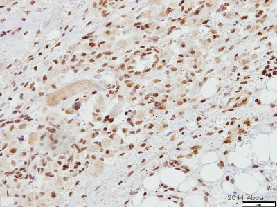
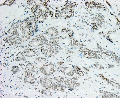 IHC image of TATA binding protein TBP staining in Human breast ductal carcinoma formalin fixed paraffin embedded tissue section, performed on a Leica BondTM system using the standard protocol F. The section was pre-treated using heat mediated antigen retrieval with sodium citrate buffer (pH6, epitope retrieval solution 1) for 20 mins. The section was then incubated with ab63766, 10µg/ml, for 15 mins at room temperature and detected using an HRP conjugated compact polymer system. DAB was used as the chromogen. The section was then counterstained with haematoxylin and mounted with DPX.For other IHC staining systems (automated and non-automated) customers should optimize variable parameters such as antigen retrieval conditions, primary antibody concentration and antibody incubation times.
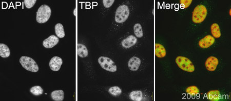 ab63766 (1/500) staining TATA binding protein (TBP) in HeLa cells (green). Cells were fixed with paraformaldehyde, permeabilised with 0.5% Triton X100/PBS and counterstained with DAPI (red) in order to highlight the nucleus. Please refer to abreview for further experimental details.See Abreview
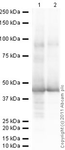
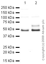 All lanes : Anti-TATA binding protein TBP antibody - Nuclear Loading Control and ChIP Grade (ab63766) at 1 µg/mlLane 1 : HeLa Whole Cell LysateLane 2 : HepG2 (Human hepatocellular liver carcinoma cell line) Whole Cell LysateLysates/proteins at 10 µg per lane.SecondaryGoat polyclonal to Rabbit IgG - H&L - Pre-Adsorbed (HRP) at 1/3000 dilutionPerformed under reducing conditions.
Product References
Microglia-induced IL-6 protects against neuronal loss following HSV-1 infection - Microglia-induced IL-6 protects against neuronal loss following HSV-1 infection
Chucair-Elliott AJ, Conrady C, Zheng M, Kroll CM, Lane TE, Carr DJ. Glia. 2014 Sep;62(9):1418-34.
GnRH increases c-Fos half-life contributing to higher FSHbeta induction. - GnRH increases c-Fos half-life contributing to higher FSHbeta induction.
Reddy GR, Xie C, Lindaman LL, Coss D. Mol Endocrinol. 2013 Feb;27(2):253-65.
A novel monoclonal antibody to secreted frizzled-related protein 2 inhibits tumor - A novel monoclonal antibody to secreted frizzled-related protein 2 inhibits tumor
Fontenot E, Rossi E, Mumper R, Snyder S, Siamakpour-Reihani S, Ma P, Hilliard E, Bone B, Ketelsen D, Santos C, Patterson C, Klauber-DeMore N. Mol Cancer Ther. 2013 May;12(5):685-95.
Nuclear translocation of B-cell-specific transcription factor, BACH2, modulates - Nuclear translocation of B-cell-specific transcription factor, BACH2, modulates
Chen Z, Pittman EF, Romaguera J, Fayad L, Wang M, Neelapu SS, McLaughlin P, Kwak L, McCarty N. PLoS One. 2013 Aug 2;8(8):e69126.
Differential regulation of osteoclastogenesis by Notch2/Delta-like 1 and - Differential regulation of osteoclastogenesis by Notch2/Delta-like 1 and
Sekine C, Koyanagi A, Koyama N, Hozumi K, Chiba S, Yagita H. Arthritis Res Ther. 2012 Mar 5;14(2):R45.
Transcriptional suppression of Sertoli cell Timp2 in rodents following - Transcriptional suppression of Sertoli cell Timp2 in rodents following
Yao PL, Lin YC, Richburg JH. Biol Reprod. 2011 Dec;85(6):1203-15.