Anti-Thioredoxin / TRX antibody
| Name | Anti-Thioredoxin / TRX antibody |
|---|---|
| Supplier | Abcam |
| Catalog | ab26320 |
| Prices | $398.00 |
| Sizes | 100 µg |
| Host | Rabbit |
| Clonality | Polyclonal |
| Isotype | IgG |
| Applications | IP WB ICC/IF ICC/IF IHC-P |
| Species Reactivities | Mouse, Cat, Human, Rat, Sheep, Bovine, Pig |
| Antigen | Synthetic peptide conjugated to KLH derived from within residues 50 to the C-terminus of Human Thioredoxin/ TRX |
| Description | Rabbit Polyclonal |
| Gene | TXN |
| Conjugate | Unconjugated |
| Supplier Page | Shop |
Product images
![Thioredoxin / TRX was immunoprecipitated using 0.5mg Hela whole cell extract, 5µg of Rabbit polyclonal to Thioredoxin / TRX and 50µl of protein G magnetic beads (+). No antibody was added to the control (-).The antibody was incubated under agitation with Protein G beads for 10min, Hela whole cell extract lysate diluted in RIPA buffer was added to each sample and incubated for a further 10min under agitation.Proteins were eluted by addition of 40µl SDS loading buffer and incubated for 10min at 70°C; 10µl of each sample was separated on a SDS PAGE gel, transferred to a nitrocellulose membrane, blocked with 5% BSA and probed with ab26320.Secondary: Mouse monoclonal [SB62a] Secondary Antibody to Rabbit IgG light chain (HRP) (ab99697).Band: 12kDa; Thioredoxin / TRX](http://www.bioprodhub.com/system/product_images/ab_products/2/sub_5/10510_ab26320-196689-IPV022ab2632020mMod.jpg) Thioredoxin / TRX was immunoprecipitated using 0.5mg Hela whole cell extract, 5µg of Rabbit polyclonal to Thioredoxin / TRX and 50µl of protein G magnetic beads (+). No antibody was added to the control (-).The antibody was incubated under agitation with Protein G beads for 10min, Hela whole cell extract lysate diluted in RIPA buffer was added to each sample and incubated for a further 10min under agitation.Proteins were eluted by addition of 40µl SDS loading buffer and incubated for 10min at 70°C; 10µl of each sample was separated on a SDS PAGE gel, transferred to a nitrocellulose membrane, blocked with 5% BSA and probed with ab26320.Secondary: Mouse monoclonal [SB62a] Secondary Antibody to Rabbit IgG light chain (HRP) (ab99697).Band: 12kDa; Thioredoxin / TRX
Thioredoxin / TRX was immunoprecipitated using 0.5mg Hela whole cell extract, 5µg of Rabbit polyclonal to Thioredoxin / TRX and 50µl of protein G magnetic beads (+). No antibody was added to the control (-).The antibody was incubated under agitation with Protein G beads for 10min, Hela whole cell extract lysate diluted in RIPA buffer was added to each sample and incubated for a further 10min under agitation.Proteins were eluted by addition of 40µl SDS loading buffer and incubated for 10min at 70°C; 10µl of each sample was separated on a SDS PAGE gel, transferred to a nitrocellulose membrane, blocked with 5% BSA and probed with ab26320.Secondary: Mouse monoclonal [SB62a] Secondary Antibody to Rabbit IgG light chain (HRP) (ab99697).Band: 12kDa; Thioredoxin / TRX
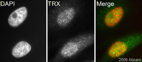 ab26320, at 1/200 dilution, staining human TRX in assynchronous HeLa cells by ICC/IF.See Abreview
![Immunofluorescent detection of Thioredoxin /TRX using antibody ab26320. Chinese Hamster Cell (Ovary cells) were fixed in formaldehyde and permeabilized in 0.1% Triton X in PBS/3% BSA. Primary antibody was incubated at 1/1000 for 1h @ 4°C in PBS containing 3% BSA. Secondary antibody: anti-rabbit conjugated to Alexa Fluor® 568 used @ 1/300. CHO cells co-transfected with plasmids encoding GFP and a mutated form of human thioredoxin which does not enter the nucleus, and labelled using ab26320 (1/1000) and Alexa 568 (1/300). Panel [A] demonstrates GFP immunoreactivity. [B ]thioredoxin immunoreactity; many of the same cells are immunoreactive for both GFP and thioredoxin. Panel [D] thioredoxin primary antibody was omitted, hence there is no immunoeactivity. See Abreview](http://www.bioprodhub.com/system/product_images/ab_products/2/sub_5/10512_Thioredoxin-TRX-Primary-antibodies-ab26320-2.jpg) Immunofluorescent detection of Thioredoxin /TRX using antibody ab26320. Chinese Hamster Cell (Ovary cells) were fixed in formaldehyde and permeabilized in 0.1% Triton X in PBS/3% BSA. Primary antibody was incubated at 1/1000 for 1h @ 4°C in PBS containing 3% BSA. Secondary antibody: anti-rabbit conjugated to Alexa Fluor® 568 used @ 1/300. CHO cells co-transfected with plasmids encoding GFP and a mutated form of human thioredoxin which does not enter the nucleus, and labelled using ab26320 (1/1000) and Alexa 568 (1/300). Panel [A] demonstrates GFP immunoreactivity. [B ]thioredoxin immunoreactity; many of the same cells are immunoreactive for both GFP and thioredoxin. Panel [D] thioredoxin primary antibody was omitted, hence there is no immunoeactivity. See Abreview
Immunofluorescent detection of Thioredoxin /TRX using antibody ab26320. Chinese Hamster Cell (Ovary cells) were fixed in formaldehyde and permeabilized in 0.1% Triton X in PBS/3% BSA. Primary antibody was incubated at 1/1000 for 1h @ 4°C in PBS containing 3% BSA. Secondary antibody: anti-rabbit conjugated to Alexa Fluor® 568 used @ 1/300. CHO cells co-transfected with plasmids encoding GFP and a mutated form of human thioredoxin which does not enter the nucleus, and labelled using ab26320 (1/1000) and Alexa 568 (1/300). Panel [A] demonstrates GFP immunoreactivity. [B ]thioredoxin immunoreactity; many of the same cells are immunoreactive for both GFP and thioredoxin. Panel [D] thioredoxin primary antibody was omitted, hence there is no immunoeactivity. See Abreview
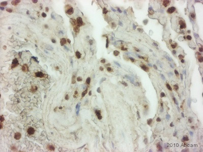 ab26320 staining Thioredoxin/TRX in Human lung tissue sections by IHC-P (Formaldehyde-fixed, paraffin-embedded sections). Tissue was fixed with formaldehyde and blocked with 5 minutes of peroxidase block followed by 10 minutes of protein block at 20°C; antigen retrieval was heat mediated in retrieval solution. Samples were incubated with primary antibody (1/2000 in antibody diluent) for 45 minutes at 20°C. An undiluted HRP-conjugated polymer goat anti-mouse/rabbit IgG polyclonal polymer was used as the secondary antibody.See Abreview
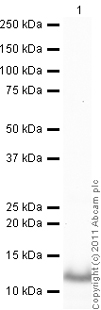 developed using the ECL techniquePerformed under reducing conditions.
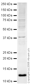 Anti-Thioredoxin / TRX antibody (ab26320) at 1 µg/ml + HeLa whole cell lysate at 20 µgSecondaryGoat polyclonal to Rabbit IgG (Alexa Fluor® 680) at 1/10000 dilutionPerformed under reducing conditions.
Product References
Increased carbonylation of the lipid phosphatase PTEN contributes to Akt2 - Increased carbonylation of the lipid phosphatase PTEN contributes to Akt2
Shearn CT, Smathers RL, Backos DS, Reigan P, Orlicky DJ, Petersen DR. Free Radic Biol Med. 2013 Dec;65:680-92. doi:
Proteomic analysis of human blastocoel fluid and blastocyst cells. - Proteomic analysis of human blastocoel fluid and blastocyst cells.
Jensen PL, Beck HC, Petersen J, Hreinsson J, Wanggren K, Laursen SB, Sorensen PD, Christensen ST, Andersen CY. Stem Cells Dev. 2013 Apr 1;22(7):1126-35.
Novel potential interacting partners of fibronectin in spontaneous animal model - Novel potential interacting partners of fibronectin in spontaneous animal model
Treutlein G, Dorsch R, Euler KN, Hauck SM, Amann B, Hartmann K, Deeg CA. PLoS One. 2012;7(12):e51391.
Up-regulation of p27(kip1) contributes to Nrf2-mediated protection against - Up-regulation of p27(kip1) contributes to Nrf2-mediated protection against
Li J, Zhang C, Xing Y, Janicki JS, Yamamoto M, Wang XL, Tang DQ, Cui T. Cardiovasc Res. 2011 May 1;90(2):315-24.
Thioredoxin-interacting protein links oxidative stress to inflammasome - Thioredoxin-interacting protein links oxidative stress to inflammasome
Zhou R, Tardivel A, Thorens B, Choi I, Tschopp J. Nat Immunol. 2010 Feb;11(2):136-40.
TXNL6 is a novel oxidative stress-induced reducing system for methionine - TXNL6 is a novel oxidative stress-induced reducing system for methionine
Brennan LA, Lee W, Kantorow M. PLoS One. 2010 Nov 4;5(11):e15421.