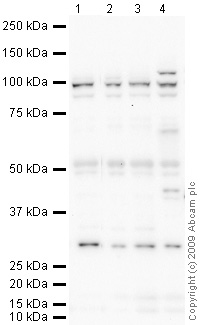
All lanes : Anti-TNPO3 antibody (ab71388) at 1 µg/mlLane 1 : Jurkat Whole Cell Lysate - Staurosporine Treated (24hr, 500nM)Lane 2 : Y79 (Human retinoblastoma cell line) Whole Cell Lysate Lane 3 : Ramos (Human Burkitt's lymphoma cell line) Whole Cell Lysate Lane 4 : MEF1 (Mouse embryonic fibroblast cell line) Whole Cell Lysate Lysates/proteins at 10 µg per lane.SecondaryGoat polyclonal to Rabbit IgG - H&L - Pre-Adsorbed (HRP) at 1/3000 dilutionPerformed under reducing conditions.
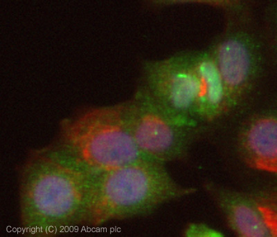
ICC/IF image of ab71388 stained MCF7 cells. The cells were 4% paraformaldehyde fixed (10 min) and then incubated in 1%BSA / 10% normal goat serum / 0.3M glycine in 0.1% PBS-Tween for 1h to permeabilise the cells and block non-specific protein-protein interactions. The cells were then incubated with the antibody (ab71388, 5µg/ml) overnight at +4°C. The secondary antibody (green) was ab96899, DyLight® 488 goat anti-rabbit IgG (H+L) used at a 1/250 dilution for 1h. Alexa Fluor® 594 WGA was used to label plasma membranes (red) at a 1/200 dilution for 1h. DAPI was used to stain the cell nuclei (blue) at a concentration of 1.43µM. This antibody also gave a positive result in 4% paraformaldehyde fixed (10 min) HeLa, Hek293 and HepG2 cells at 5µg/ml.