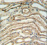
ADH7 Antibody IHC of formalin-fixed and paraffin-embedded lung tissue followed by peroxidase-conjugated secondary antibody and DAB staining.
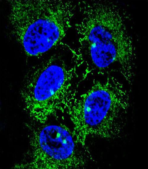
Confocal immunofluorescent of ADH7 Antibody with NCI-H460 cell followed by Alexa Fluor 488-conjugated goat anti-rabbit lgG (green). DAPI was used to stain the cell nuclear (blue).
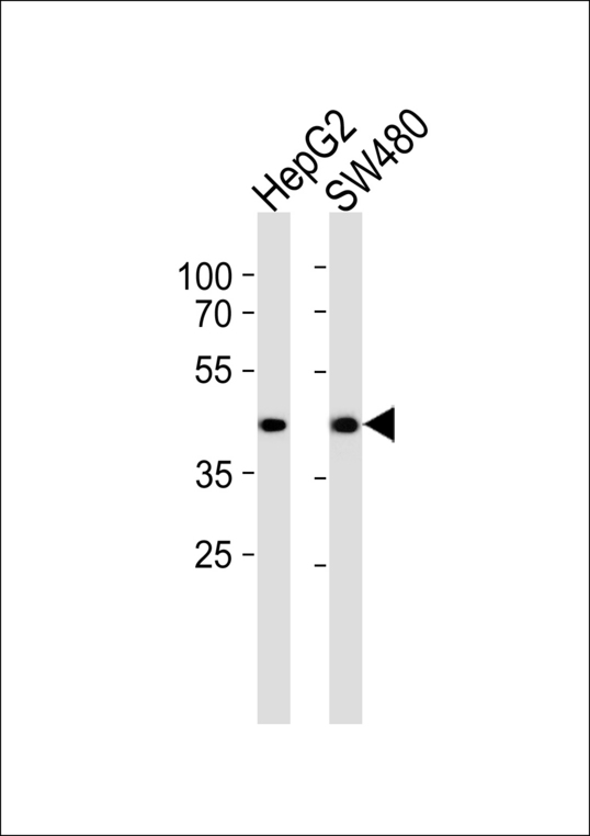
Western blot of lysates from HepG2, SW480 cell line (from left to right), using ADH7 Antibody. Antibody was diluted at 1:1000 at each lane. A goat anti-rabbit IgG H&L (HRP) at 1:5000 dilution was used as the secondary antibody. Lysates at 35ug per lane.
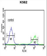
ADH7 Antibody flow cytometry of K562 cells (right histogram) compared to a negative control cell (left histogram). FITC-conjugated goat-anti-rabbit secondary antibodies were used for the analysis.