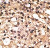
Formalin-fixed and paraffin-embedded human cancer tissue reacted with the primary antibody, which was peroxidase-conjugated to the secondary antibody, followed by DAB staining. This data demonstrates the use of this antibody for immunohistochemistry; clinical relevance has not been evaluated. BC = breast carcinoma; HC = hepatocarcinoma.
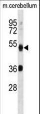
APG4B Antibody (R31) western blot of mouse cerebellum tissue lysates (35 ug/lane). The APG4B antibody detected the APG4B protein (arrow).
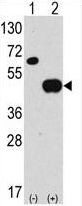
Western blot of anti-hAPG4B-R31 antibody in 293 cell line lysates transiently transfected with the ATG4B gene (2 ug/lane). hAPG4B-R31(arrow) was detected using the purified antibody.
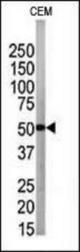
The anti-APG4B antibody is used in Western blot to detect APG4B in CEM tissue lysate