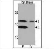
ARG54709 anti-LC3A antibody WB image Western blot: rat brain lysate stained with ARG54709 anti-LC3A antibody. Both lipidated (arrow, II) and non-lipidated APG8a (arrow, I) were detected in membrane fraction (P) but only non-lipidated LC3 was detected in soluble fraction (S).
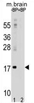
ARG54709 anti-LC3A antibody WB image Western blot: 35 μg of mouse brain tissue lysates stained with ARG54709 anti-LC3A antibody.
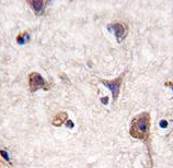
ARG54709 anti-LC3A antibody IHC image Immunohistochemistry: human brain tissue stained with ARG54709 anti-LC3A antibody.
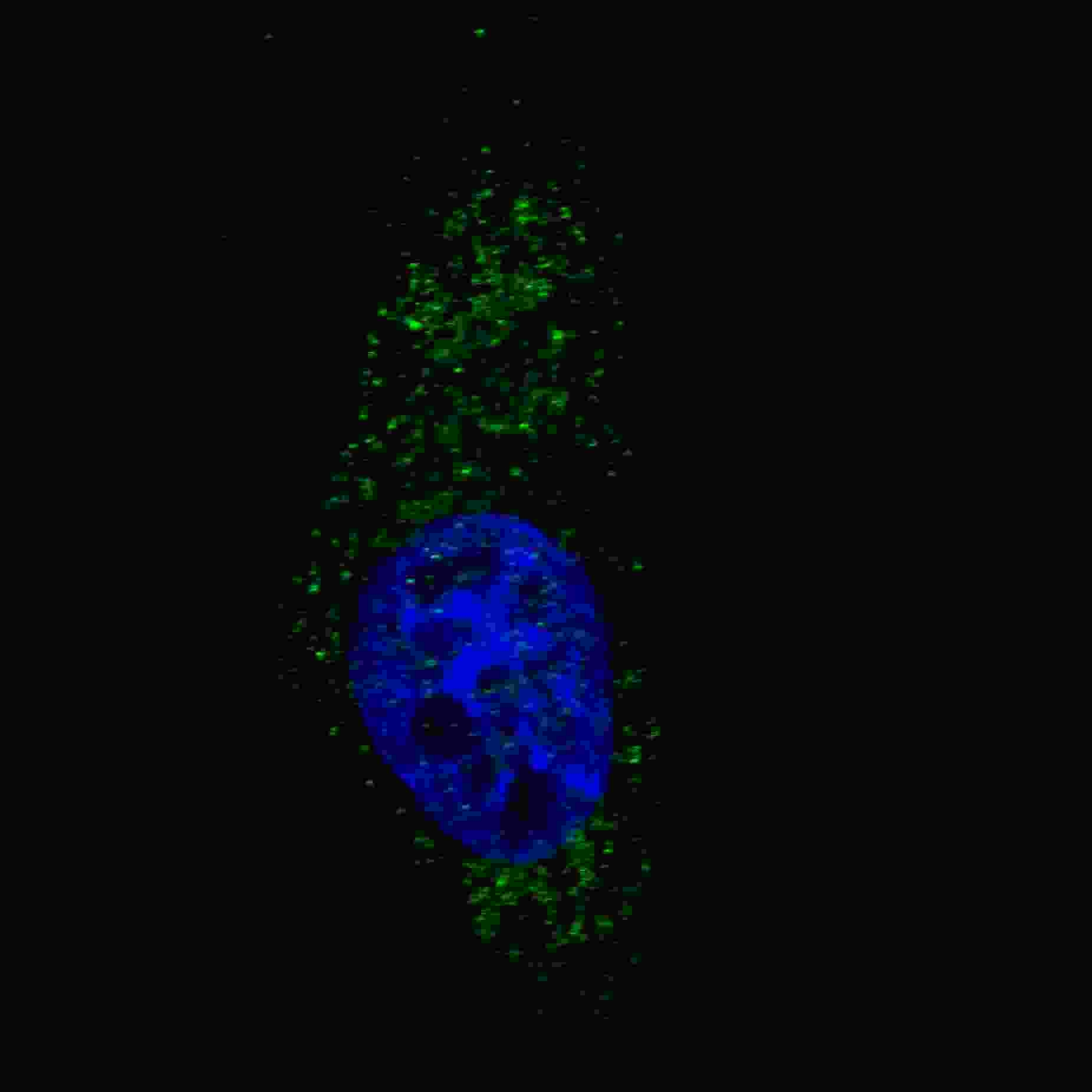
ARG54709 anti-LC3A antibody IF image Immunofluorescence: U251 cells stained with ARG54709 anti-LC3A antibody. U251 cells were treated with Chloroquine (50 μM,16h), then fixed with 4% PFA (20 min), permeabilized with Triton X-100 (0.2%, 30 min). Cells were then incubated with AP1801a LC3 (APG8A) (N-term) primary antibody (1:200, 2 h at room temperature). Nuclei were counterstained with Hoechst 33342 (blue) (10 μg/ml, 5 min). LC3 immunoreactivity is localized to autophagic vacuoles in the cytoplasm of U251 cells.