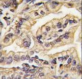
Formalin-fixed and paraffin-embedded human lung carcinoma tissue reacted with CSF1R Antibody , which was peroxidase-conjugated to the secondary antibody, followed by DAB staining. This data demonstrates the use of this antibody for immunohistochemistry; clinical relevance has not been evaluated.
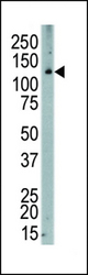
Western blot of anti-CSF1R antibody in human placenta. CSF1R (arrow) was detected using purified antibody. Secondary HRP-anti-rabbit was used for signal visualization with chemiluminescence.
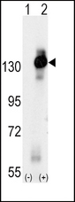
Western blot of CSF1R (arrow) using rabbit polyclonal CSF1R Antibody. 293 cell lysates (2 ug/lane) either nontransfected (Lane 1) or transiently transfected with the CSF1R gene (Lane 2) (Origene Technologies).
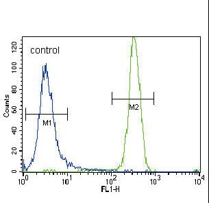
MCSF Receptor (CSF1R) Antibody flow cytometry of NCI-H460 cells (right histogram) compared to a negative control cell (left histogram). FITC-conjugated goat-anti-rabbit secondary antibodies were used for the analysis.