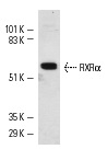
RXRα (D-20): sc-553. Western blot analysis of RXRα expression in HeLa nuclear extracts.
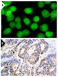
RXRα (D-20): sc-553. Immunofluorescence staining of methanol-fixed HeLa cells (A) and immunoperoxidase staining of formalin-fixed, paraffin-embedded human colon carcinoma tissue (B) showing nuclear staining.
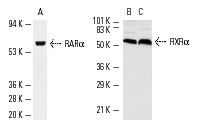
Western blot analysis of RARα (A) and RXRα (B,C) expression in HeLa (A) nuclear extract and NIH/3T3 (B) and KNRK (C) whole cell lysates. Antibodies tested include RARα (C-20): sc-551 (A) and RXRα (D-20): sc-553 (B,C).
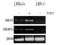
RXRα (D-20): sc-553. ChIP analysis of LXR dependent promoter recruitment of RXR in bone marrow-derived macrophages from LXR+/+ and LXR-/- mice in response to the LXR ligand T1317. Data kindly provided by M.G. Rosenfeld and reproduced with permission from Wagner et al., Mol. Cell. Biol. 2003, 23:5780-5789.
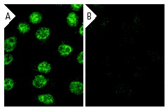
RXRα siRNA (h): sc-36447. Immunofluorescence staining of methanol-fixed, control HeLa (A) and RXRα siRNA silenced HeLa (B) cells showing diminished nuclear staining in the siRNA silenced cells. Cells probed with RXRα (D-20): sc-553.
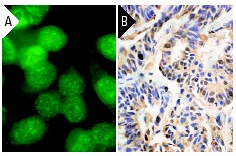
RXRα (D-20): sc-553. Immunofluorescence staining of methanol-fixed HeLa cells (A) and immunoperoxidase staining of formalin-fixed, paraffin-embedded human colon carcinoma tissue (B) showing nuclear staining.
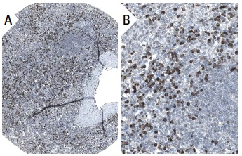
RXRα (D-20): sc-553. Immunoperoxidase staining of formalin fixed, paraffin-embedded human spleen tissue showing nuclear staining of cells in red pulp (low and high magnification). Kindly provided by The Swedish Human Protein Atlas (HPA) program.
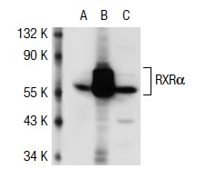
RXRα (D-20): sc-553. Western blot analysis of RXRα expression in non-transfected 293T: sc-117752 (A), human RXRα transfected 293T: sc-111936 (B) and K-562 (C) whole cell lysates.