Anti-Amphiphysin II Antibody
| Name | Anti-Amphiphysin II Antibody |
|---|---|
| Supplier | Sigma-Aldrich |
| Catalog | ABT21 |
| Prices | $319.00 |
| Sizes | abt21 |
| Clonality | Polyclonal |
| Isotype | IgG |
| Applications | WB |
| Species Reactivities | Mouse, Rat |
| Purity/Format | Affinity Purfied |
| Description | Polyclonal |
| Gene | Bin1 |
| Supplier Page | Shop |
Product images
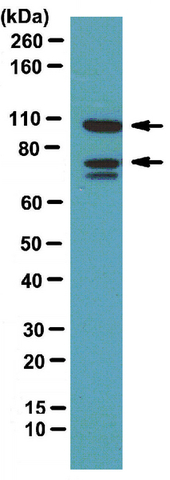 Western Blotting Analysis:
Representative lot data.
Mouse brain tissue lysate was probed with Anti-Amphiphysin II (1:1000 dilution).
Proteins were visualized using a Donkey Anti-Rabbit IgG secondary antibody conjugated to HRP (Cat. No. AP182P) and detected using a chemiluminescence detection system (Cat. No. 17-373).
Arrows indicate Amphiphysin I and II (~ 105 kDa, 75 kDa).
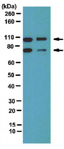 Western Blotting Analysis:
 Representative lot data.
Rat brain membrane (lane 1) and mouse brain membrane (lane 2) tissue lysates were probed with Anti-Amphiphysin II (1:1000 dilution).
Proteins were visualized using a Donkey Anti-Rabbit IgG secondary antibody conjugated to HRP (Cat. No. AP182P) and detected using a chemiluminescence detection system (Cat. No. 17-373).
Arrows indicate Amphiphysin I and II (~ 105 kDa, 75 kDa).