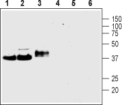
Western blot analysis of rat brain membranes (lanes 1 and 4), mouse brain membranes (lanes 2 and 5) and rat PC12 pheochromocytoma cell line lysate (lanes 3 and 6): 1-3.Anti-Synaptophysinantibody (#AG1535), (1:400).4-6. Anti-Synaptophysin antibody, preincubated with the control peptide antigen.

Expression of Synaptophysin in rat DRG Immunohistochemical staining of rat dorsal root ganglia (DRG) frozen sections using Anti-Synaptophysin antibody (#AG1535), (1:100). Synaptophysin (red) is expressed in DRG neurons. Hoechst 33342 (blue) shows nuclear staining and is used as the counterstain.
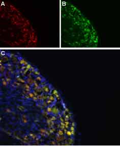
Colocalization of NaV1.8 and Synaptophysin in rat DRG Immunohistochemical staining of rat DRG frozen section using Anti-NaV1.8-ATTO-594 antibody (#ASC-016-AR) and Anti-Synaptophysin antibody (#AG1535). A. NaV1.8 staining (red). B. Synaptophysin staining (green). C. Merged image demonstrates a partial overlap in the distribution of Nav1.8 and Synaptophysin within the DRGs. DAPI is used as the counterstain (blue).