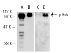
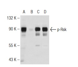
Western blot analysis of λ phosphatase-treated (A,C) and untreated (B,D) rat recombinant Rsk-1. Antibodies tested include Rsk-1 (C-21): sc-231 (A,B) and p-Rsk-1/2 (Thr 359/Ser 363)-R: sc-12898-R (C,D).
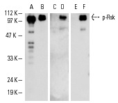
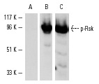
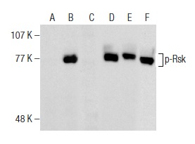
Western blot analysis of λ phosphatase-treated (A,C,E) and untreated (B,D,F) active human recombinant Rsk. Antibodies tested include Rsk-1 (C-21): sc-231 (A,B), p-Rsk (Ser 376): sc-12883 (C,D) and p-Rsk (Ser 376)-R: sc-12883-R (E,F).
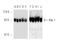
Western blot analysis of Rsk-1 expression in MDCK (A,F), Mv 1 Lu (B,G), A-431 (C,H), HeLa (D,I) and MCF7 (E,J) whole cell lysates. Antibodies tested include Rsk-1 (C-21): sc-231 (A-E) and Rsk-1 (C-21)-G: sc-231-G (F-J).
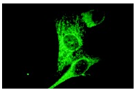
Rsk-1 (C-21): sc-231. Cytoplasmic immunofluorescence staining of methanol-fixed NIH/3T3 cells.
Western blot analysis of Rsk-1/2/4 phosphorylation in HeLa whole cell lysate (A-C). Blots were probed with p-Rsk-1/2/4 (Ser 363)-R: sc-17033-R preincubated with cognate phosphorylated (A) and unphosphorylated (B) peptide and Rsk-1 (C-21): sc-231 (C).
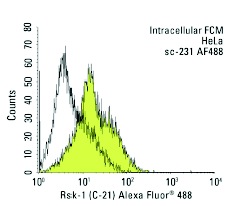
Rsk-1 (C-21) Alexa Fluor 488: sc-231 AF488. Intracellular FCM analysis of fixed and permeabilized HeLa cells. Black line histogram represents the isotype control, normal rabbit IgG: sc-45068.
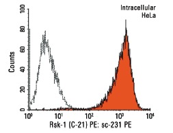
Rsk-1 (C-21) PE: sc-231 PE. Intracellular FCM analysis of fixed and permeabilized HeLa cells. Black line histogram represents the isotype control, normal rabbit IgG: sc-3871.
Western blot analysis of Rsk phosphorylation in untreated (A,C) and lambda protein phosphatase treated (B,D) HeLa whole cell lysates. Antibodies tested include p-Rsk (Ser 227)-R: sc-12445-R (A,B) and Rsk-1 (C-21): sc-231 (C,D).
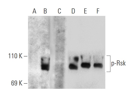
Western blot analysis of Rsk phosphorylation in untreated (A,D), serum-starved, PMA-treated (B,E), serum-starved, PMA and lambda protein phosphatase (sc-200312A) treated (C,F) HeLa whole cell lysates. Antibodies tested include p-Rsk (Ser 380)-R: sc-11756-R (A,B,C) and Rsk-1 (C-21): sc-231 (D,E,F).
Western blot analysis of Rsk phosphorylation in untreated (A,D), serum-starved, PMA-treated (B,E), serum-starved, PMA and lambda protein phosphatase (sc-200312A) treated (C,F) HeLa whole cell lysates. Antibodies tested include p-Rsk-1/2/4 (Ser 363)-R: sc-17033-R (A,B,C) and Rsk-1 (C-21): sc-231 (D,E,F).
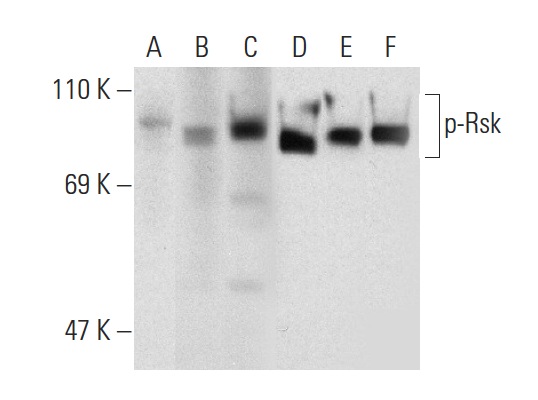
Western blot analysis of Rsk phosphorylation in untreated (A,D), serum-starved, PMA-treated (B,E), and serum-starved, PMA and lambda protein phosphatase (sc-200312A) treated (C,F) HeLa whole cell lysates. Antibodies tested include p-Rsk (Ser 227)-R: sc-12445-R (A,B,C) and Rsk-1 (C-21): sc-231 (D,E,F).
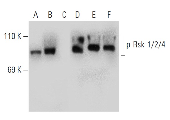
Western blot analysis of Rsk phosphorylation in untreated (A,D), serum-starved, PMA-treated (B,E), and serum-starved, PMA and lambda protein phosphatase (sc-200312A) treated (C,F) HeLa whole cell lysates. Antibodies tested include p-Rsk (Thr 359/Ser 363)-R: sc-12898-R (A,B,C) and Rsk-1 (C-21): sc-231 (D,E,F).
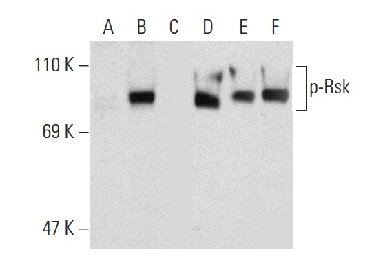
Western blot analysis of Rsk phosphorylation in untreated (A,D), PMA treated (B,E) and PMA and lambda protein phosphatase (sc-200312A) treated (C,F) HeLa whole cell lysates. Antibodies tested include p-Rsk (C-5): sc-377526 (A,B,C) and Ribosomal Rsk-1 (C-21): sc-231 (D,E,F).
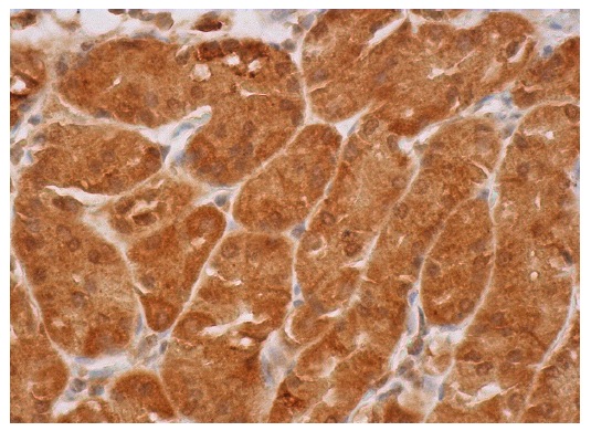
Rsk-1 (C-21)-G: sc-231-G. Immunoperoxidase staining of formalin fixed, paraffin-embedded human upper stomach tissue showing cytoplasmic and nuclear staining of glandular cells.