Anti-GCG / Glucagon Antibody (aa119-148) IHC-plusâ¢
| Name | Anti-GCG / Glucagon Antibody (aa119-148) IHC-plus⢠|
|---|---|
| Supplier | LifeSpan Bioscience |
| Catalog | LS-B10219 |
| Prices | $395.00 |
| Sizes | 200 µl |
| Host | Rabbit |
| Clonality | Polyclonal |
| Applications | IHC-P WB |
| Species Reactivities | Human |
| Antigen | GCG / Glucagon antibody was raised against kLH-conjugated synthetic peptide between amino acids 119-148 from C-terminal region of human Glucagon. |
| Purity/Format | Ammonium sulfate precipitation |
| Blocking Peptide | GCD / GCDH Antibody Blocking Peptide |
| Description | Rabbit Polyclonal |
| Gene | GCG |
| Conjugate | Unconjugated |
| Supplier Page | Shop |
Product images
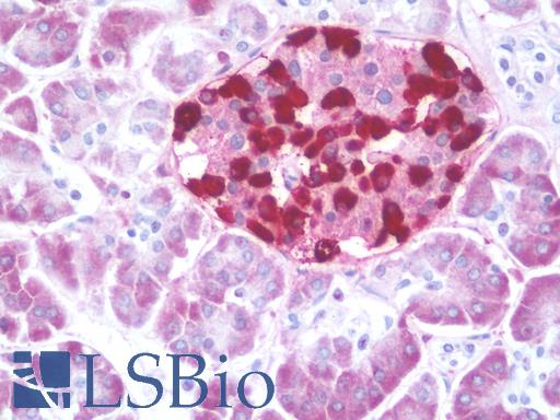 Anti-GCG / Glucagon antibody IHC staining of human pancreas. Immunohistochemistry of formalin-fixed, paraffin-embedded tissue after heat-induced antigen retrieval. Antibody LS-B10219 dilution 1:200.
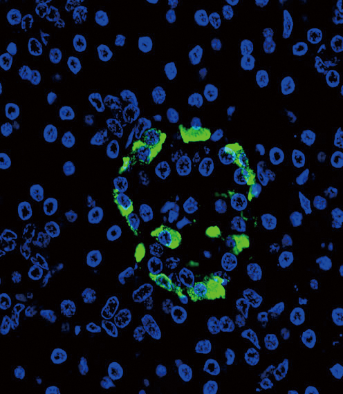 Confocal immunofluorescent of Glucagon Antibody with pancreas tissue followed by Alexa Fluor 488-conjugated goat anti-rabbit lgG (green). DAPI was used to stain the cell nuclear (blue).
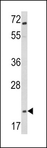 Western blot of Glucagon Antibody in Jurkat cell line lysates (35 ug/lane). GCG (arrow) was detected using the purified antibody.
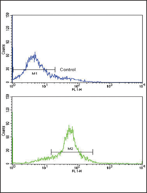 Flow cytometric of WiDr cells using Glucagon Antibody (bottom histogram) compared to a negative control cell (top histogram). FITC-conjugated goat-anti-rabbit secondary antibodies were used for the analysis.