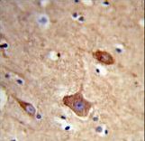
Formalin-fixed and paraffin-embedded human brain tissue reacted with NXPH1 Antibody , which was peroxidase-conjugated to the secondary antibody, followed by DAB staining. This data demonstrates the use of this antibody for immunohistochemistry; clinical relevance has not been evaluated.
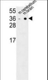
NXPH1 Antibody western blot of mouse cerebellum,brain tissue lysates (35 ug/lane). The NXPH1 antibody detected the NXPH1 protein (arrow).
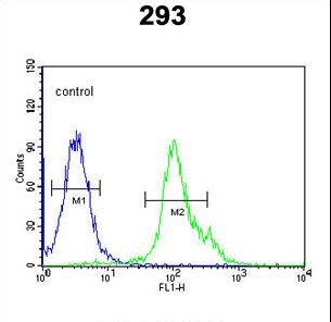
NXPH1 Antibody flow cytometry of 293 cells (right histogram) compared to a negative control cell (left histogram). FITC-conjugated goat-anti-rabbit secondary antibodies were used for the analysis.