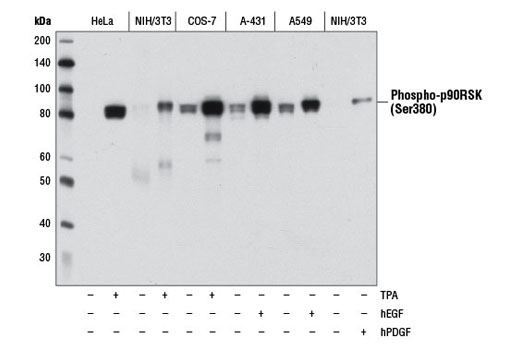
Western blot analysis of extracts from various cell lines, starved overnight and either untreated (-) or treated (+) with TPA #4174 (200 nM, 15 min), Human Epidermal Growth Factor (hEGF) #8916 (100 ng/mL, 15 min), or Human Platelet-Derived Growth Factor BB (hPDGF-BB) #8912 (100 ng/mL, 15 min) as indicated, using Phospho-p90RSK (Ser380) (D3H11) Rabbit mAb.
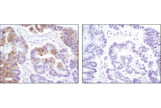
Immunohistochemical analysis of paraffin-embedded human colon carcinoma, control (left) or λ phosphatase-treated (right), using Phospho-p90RSK (Ser380) (D3H11) Rabbit mAb.
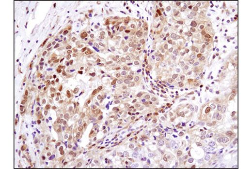
Immunohistochemical analysis of paraffin-embedded human ovarian carcinoma using Phospho-p90RSK (Ser380) (D3H11) Rabbit mAb.
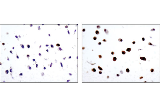
Immunohistochemical analysis of paraffin-embedded HeLa cell pellets, untreated (left) or treated with TPA #4174 (right), using Phospho-p90RSK (Ser380) (D3H11) Rabbit mAb.
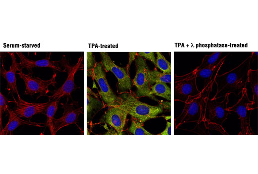
Confocal immunofluorescent analysis of NIH/3T3 cells, serum-starved (left), treated with TPA #4174 (200 nM, 15 min; center), or treated with TPA followed by λ phosphatase (right), using Phospho-p90RSK (Ser380) (D3H11) Rabbit mAb (green). Actin filaments were labeled with DY-554 phalloidin (red). Blue pseudocolor = DRAQ5 ® #4084 (fluorescent DNA dye).