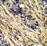
Formalin-fixed and paraffin-embedded human colon carcinoma reacted with YWHAG Antibody , which was peroxidase-conjugated to the secondary antibody, followed by DAB staining. This data demonstrates the use of this antibody for immunohistochemistry; clinical relevance has not been evaluated.
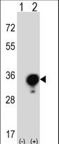
Western blot of YWHAG (arrow) using rabbit polyclonal YWHAG Antibody. 293 cell lysates (2 ug/lane) either nontransfected (Lane 1) or transiently transfected (Lane 2) with the YWHAG gene.
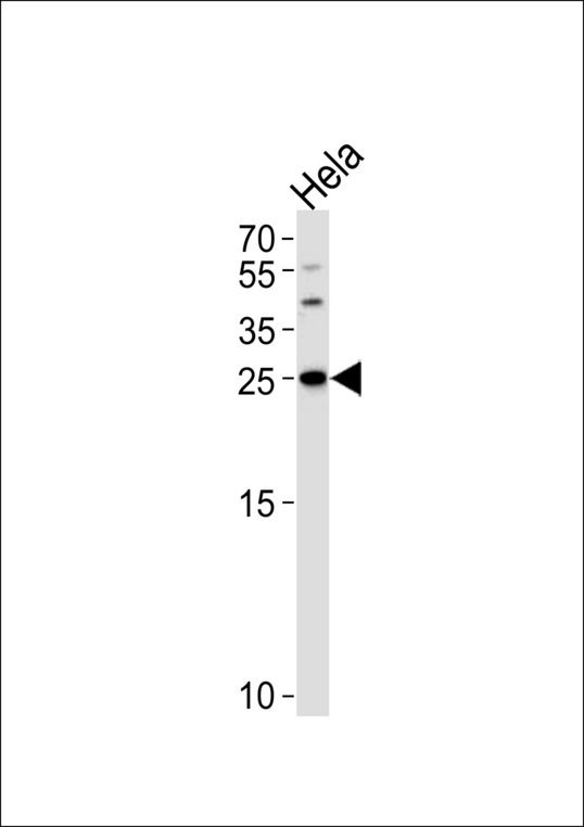
Western blot of lysate from HeLa cell line, using YWHAG Antibody. Antibody was diluted at 1:1000 at each lane. A goat anti-rabbit IgG H&L (HRP) at 1:5000 dilution was used as the secondary antibody. Lysate at 35ug.
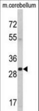
Western blot of YWHAG Antibody in mouse cerebellum tissue lysates (35 ug/lane). YWHAG (arrow) was detected using the purified antibody.
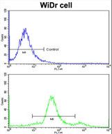
Flow cytometric of widr cells using YWHAG Antibody (bottom histogram) compared to a negative control cell (top histogram)FITC-conjugated goat-anti-rabbit secondary antibodies were used for the analysis.