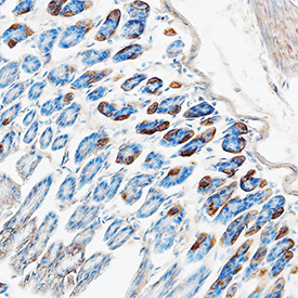
CCL11/Eotaxin was detected in perfusion fixed frozen sections of mouse colon using Goat Anti-Mouse CCL11/Eotaxin Antigen Affinity-purified Polyclonal Antibody (Catalog # AF-420-NA) at 15 µg/mL overnight at 4 °C. Tissue was stained using the Anti-Goat HRP-DAB Cell & Tissue Staining Kit (brown; Catalog # CTS008) and counterstained with hematoxylin (blue). Specific staining was localized to cytoplasm. View our protocol for Chromogenic IHC Staining of Frozen Tissue Sections.

Recombinant Mouse CCL11/Eotaxin (Catalog # 420‑ME) chemoattracts the BaF3 mouse pro‑B cell line transfected with mouse CCR3 in a dose-dependent manner (orange line). The amount of cells that migrated through to the lower chemotaxis chamber was measured by Resazurin (Catalog # AR002). Chemotaxis elicited by Recombinant Mouse CCL11/Eotaxin (10 ng/mL) is neutralized (green line) by increasing concentrations of Goat Anti-Mouse CCL11/Eotaxin Antigen Affinity-purified Polyclonal Antibody (Catalog # AF-420-NA). The ND50 is typically0.1‑0.5 µg/mL.
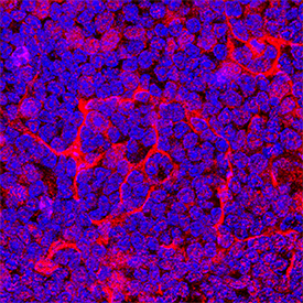
CCL11/Eotaxin was detected in perfusion fixed frozen sections of mouse thymus using Goat Anti-Mouse CCL11/Eotaxin Antigen Affinity-purified Polyclonal Antibody (Catalog # AF-420-NA) at 5 µg/mL overnight at 4 °C. Tissue was stained using the NorthernLights™ 557-conjugated Anti-Goat IgG Secondary Antibody (red; Catalog # NL001) and counterstained with DAPI (blue). Specific staining was localized to cytoplasm. View our protocol for Fluorescent IHC Staining of Frozen Tissue Sections.