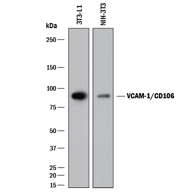
Western blot shows lysates of 3T3‑L1 mouse embryonic fibroblast adipose-like cell line and NIH-3T3 mouse embryonic fibroblast cell line. PVDF membrane was probed with 0.25 µg/mL of Goat Anti-Mouse VCAM‑1/CD106 Antigen Affinity-purified Polyclonal Antibody (Catalog # AF643) followed by HRP-conjugated Anti-Goat IgG Secondary Antibody (Catalog # HAF017). A specific band was detected for VCAM‑1/CD106 at approximately 95 kDa (as indicated). This experiment was conducted under reducing conditions and using Immunoblot Buffer Group 1.
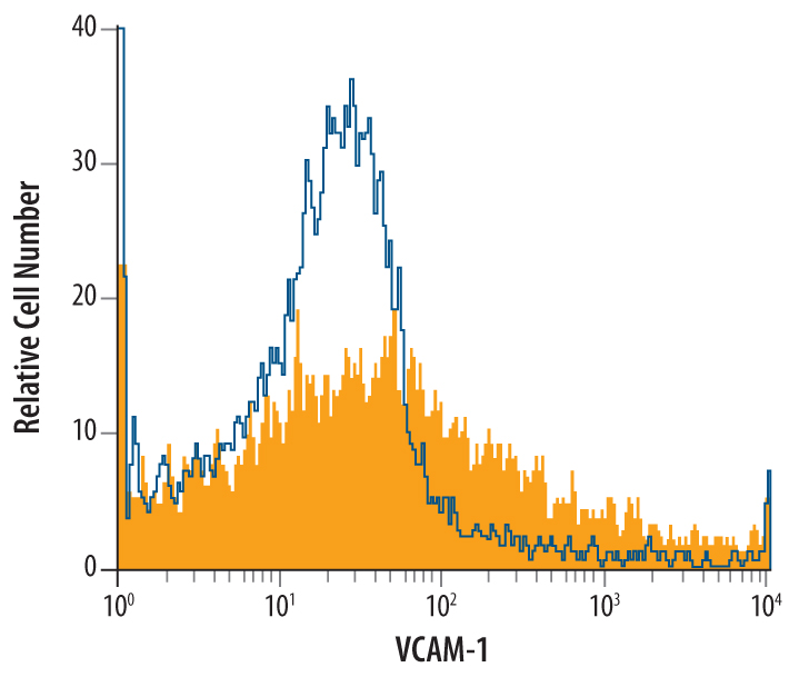
Mouse bone marrow cells were stained with Goat Anti-Mouse VCAM‑1/CD106 Antigen Affinity-purified Polyclonal Antibody (Catalog # AF643, filled histogram) or control antibody (Catalog # AB-108-C, open histogram), followed by Phycoerythrin-conjugated Anti-Goat IgG Secondary Antibody (Catalog # F0107).
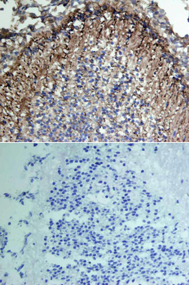
VCAM‑1/CD106 was detected in immersion fixed frozen sections of mouse embryo using Goat Anti-Mouse VCAM‑1/CD106 Antigen Affinity-purified Polyclonal Antibody (Catalog # AF643) at 15 µg/mL overnight at 4 °C. Tissue was stained using the Anti-Goat HRP-DAB Cell & Tissue Staining Kit (brown; Catalog # CTS008) and counterstained with hematoxylin (blue). Lower panel shows a lack of labeling if primary antibodies are omitted and tissue is stained only with secondary antibody followed by incubation with detection reagents. View our protocol for Chromogenic IHC Staining of Frozen Tissue Sections.
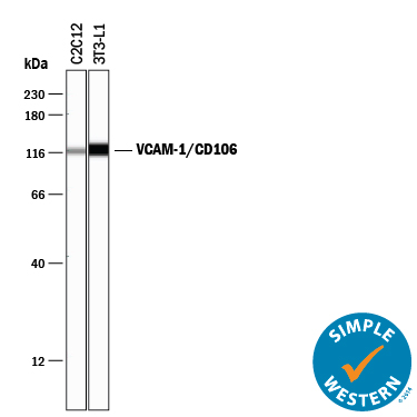
Simple Western lane view shows lysates of C2C12 mouse myoblast cell line and 3T3‑L1 mouse embryonic fibroblast adipose-like cell line, loaded at 0.2 mg/mL. A specific band was detected for VCAM‑1/CD106 at approximately 116 kDa (as indicated) using 1 µg/mL of Goat Anti-Mouse VCAM‑1/CD106 Antigen Affinity-purified Polyclonal Antibody (Catalog # AF643) followed by 1:50 dilution of HRP-conjugated Anti-Goat IgG Secondary Antibody (Catalog # HAF109). This experiment was conducted under reducing conditions and using the 12-230 kDa separation system.