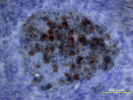
Oncostatin M/OSM was detected in immersion fixed frozen sections of mouse embryo using Mouse Oncostatin M/OSM Antigen Affinity-purified Polyclonal Antibody (Catalog # AF-495-NA) at 15 µg/mL overnight at 4 °C. Tissue was stained using the Anti-Goat HRP-DAB Cell & Tissue Staining Kit (brown; Catalog # CTS008) and counterstained with hematoxylin (blue). View our protocol for Chromogenic IHC Staining of Frozen Tissue Sections.

Oncostatin M/OSM was detected in immersion fixed frozen sections of mouse embryo (15 d.p.c, section through spinal cord) using Mouse Oncostatin M/OSM Antigen Affinity-purified Polyclonal Antibody (Catalog # AF-495-NA) at 15 µg/mL overnight at 4 °C. Tissue was stained using the Anti-Goat HRP-DAB Cell & Tissue Staining Kit (brown; Catalog # CTS008) and counterstained with hematoxylin (blue). View our protocol for Chromogenic IHC Staining of Frozen Tissue Sections.

Recombinant Mouse Oncostatin M/OSM (Catalog # 495-MO) stimulates proliferation in the NIH‑3T3 mouse embryonic fibroblast cell line in a dose-dependent manner (orange line). Proliferation elicited by Recombinant Mouse Oncostatin M/OSM (15 ng/mL) is neutralized (green line) by increasing concentrations of Mouse Oncostatin M/OSM Antigen Affinity-purified Polyclonal Antibody (Catalog # AF-495-NA). The ND50 is typically 0.6-3.0 µg/mL.


