
Cathepsin L was detected in perfusion fixed frozen sections of mouse ovary using 15 µg/mL Goat Anti-Mouse Cathepsin L Antigen Affinity-purified Polyclonal Antibody (Catalog # AF1515) overnight at 4 °C. Tissue was stained with the Anti-Goat HRP-DAB Cell & Tissue Staining Kit (brown; Catalog # CTS008) and counterstained with hematoxylin (blue). View our protocol for Chromogenic IHC Staining of Frozen Tissue Sections.
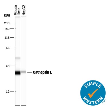
Simple Western lane view shows lysates of mouse liver tissue and HepG2 human hepatocellular carcinoma cell line, loaded at 0.2 mg/mL. Specific bands were detected for Cathepsin L at approximately 51 and 34 kDa (as indicated) using 10 µg/mL of Goat Anti-Mouse Cathepsin L Antigen Affinity-purified Polyclonal Antibody (Catalog # AF1515) followed by 1:50 dilution of HRP-conjugated Anti-Goat IgG Secondary Antibody (Catalog # HAF109). This experiment was conducted under reducing conditions and using the 12-230 kDa separation system.

Cathepsin L was detected in perfusion fixed frozen sections of mouse thymus using 15 µg/mL Goat Anti-Mouse Cathepsin L Antigen Affinity-purified Polyclonal Antibody (Catalog # AF1515) overnight at 4 °C. Tissue was stained with the Anti-Goat HRP-DAB Cell & Tissue Staining Kit (brown; Catalog # CTS0028) and counterstained with hematoxylin (blue). View our protocol for Chromogenic IHC Staining of Frozen Tissue Sections.

Western blot shows lysates of rat liver tissue, mouse liver tissue (wild type), and mouse liver tissue (knock out). PVDF membrane was probed with 1 µg/mL of Goat Anti-Mouse Cathepsin L Antigen Affinity-purified Polyclonal Antibody (Catalog # AF1515) followed by HRP-conjugated Anti-Goat IgG Secondary Antibody (Catalog # HAF017). Specific bands were detected for Cathepsin L at approximately 22-38 kDa (as indicated). This experiment was conducted under reducing conditions and using Immunoblot Buffer Group 1.