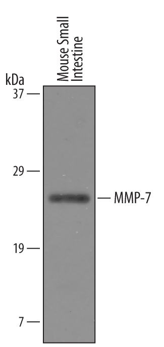
Western blot shows lysates of mouse small intestine tissue. PVDF Membrane was probed with 1 µg/mL of Goat Anti-Mouse MMP‑7 Antigen Affinity-purified Polyclonal Antibody (Catalog # AF2967) followed by HRP-conjugated Anti-Goat IgG Secondary Antibody (Catalog # HAF019). A specific band was detected for MMP‑7 at approximately 28 kDa (as indicated). This experiment was conducted under reducing conditions and using Immunoblot Buffer Group 8.
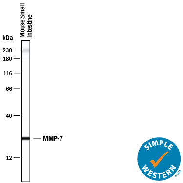
Simple Western lane view shows lysates of mouse small intestine tissue, loaded at 0.2 mg/mL. A specific band was detected for MMP‑7 at approximately 25 kDa (as indicated) using 50 µg/mL of Goat Anti-Mouse MMP‑7 Antigen Affinity-purified Polyclonal Antibody (Catalog # AF2967) followed by 1:50 dilution of HRP-conjugated Anti-Goat IgG Secondary Antibody (Catalog # HAF109). This experiment was conducted under reducing conditions and using the12-230 kDa separation system. Non-specific interaction with the 230 kDa Simple Western standard may be seen with this antibody.

MMP‑7 was detected in perfusion fixed frozen sections of mouse small intestine using Goat Anti-Mouse MMP‑7 Antigen Affinity-purified Polyclonal Antibody (Catalog # AF2967) at 15 µg/mL overnight at 4 °C. Tissue was stained using the Anti-Goat HRP-DAB Cell & Tissue Staining Kit (brown; Catalog # CTS008) and counterstained with hematoxylin (blue). Specific labeling was localized to the cytoplasm of cells in intestinal glands. View our protocol for Chromogenic IHC Staining of Frozen Tissue Sections.