
Western blot shows lysates of 3T3‑L1 mouse embryonic fibroblast adipose-like cell line. PVDF membrane was probed with 1 µg/mL of Goat Anti-Mouse Pref‑1/DLK1/FA1 Antigen Affinity-purified Polyclonal Antibody (Catalog # AF8277) followed by HRP-conjugated Anti-Goat IgG Secondary Antibody (Catalog # HAF017). A specific band was detected for Pref‑1/DLK1/FA1 at approximately 45-60 kDa (as indicated). This experiment was conducted under reducing conditions and using Immunoblot Buffer Group 1.
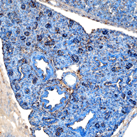
Pref‑1/DLK1/FA1 was detected in immersion fixed frozen sections of mouse embryonic lung using Goat Anti-Mouse Pref‑1/DLK1/FA1 Antigen Affinity-purified Polyclonal Antibody (Catalog # AF8277) at 1 µg/mL overnight at 4 °C. Tissue was stained using the Anti-Goat HRP-DAB Cell & Tissue Staining Kit (brown; Catalog # CTS008) and counterstained with hematoxylin (blue). Specific staining was localized to cytoplasm. View our protocol for Chromogenic IHC Staining of Frozen Tissue Sections.
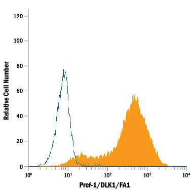
3T3‑L1 mouse embryonic fibroblast adipose-like cell line was stained with Goat Anti-Mouse Pref‑1/DLK1/FA1 Antigen Affinity-purified Polyclonal Antibody (Catalog # AF8277, filled histogram) or control antibody (Catalog # AB-108-C, open histogram), followed by Phycoerythrin-conjugated Anti-Goat IgG Secondary Antibody (Catalog # F0107).