
Western blot shows lysates of mouse spleen tissue and mouse brain tissue. PVDF membrane was probed with 1 µg/mL of Sheep Anti-Mouse IL‑34 Antigen Affinity-purified Polyclonal Antibody (Catalog # AF5195) followed by HRP-conjugated Anti-Sheep IgG Secondary Antibody (Catalog # HAF016). A specific band was detected for IL‑34 at approximately 33 kDa (as indicated). This experiment was conducted under reducing conditions and using Immunoblot Buffer Group 8.
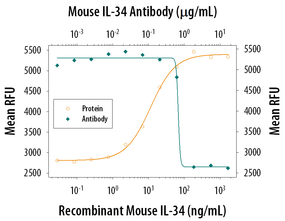
Recombinant Mouse IL‑34 (Catalog # 5195-ML) induces proliferation in the M‑NFS‑60 mouse myelogenous leukemia lymphoblast cell line in a dose-dependent manner (orange line). Proliferation elicited by Recombinant Mouse IL‑34 (100 ng/mL) is neutralized (green line) by increasing concentrations of Sheep Anti-Mouse IL‑34 Antigen Affinity-purified Polyclonal Antibody (Catalog # AF5195). The ND50 is typically 0.3-1.5 μg/mL.