
Western blot shows lysates of human cartilage tissue. PVDF membrane was probed with 1 µg/mL of Sheep Anti-Human CRTAC1 Isoform 1 Antigen Affinity-purified Polyclonal Antibody (Catalog # AF5234) followed by HRP-conjugated Anti-Sheep IgG Secondary Antibody (Catalog # HAF016). A specific band was detected for CRTAC1 at approximately 95 kDa (as indicated). This experiment was conducted under reducing conditions and using Immunoblot Buffer Group 8.
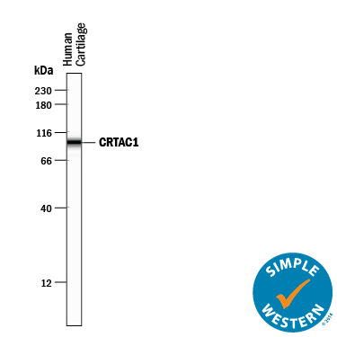
Simple Western lane view shows lysates of human cartilage tissue, loaded at 0.5 mg/mL. A specific band was detected for CRTAC1 at approximately 97 kDa (as indicated) using 10 µg/mL of Sheep Anti-Human CRTAC1 Isoform 1 Antigen Affinity-purified Polyclonal Antibody (Catalog # AF5234) followed by 1:50 dilution of HRP-conjugated Anti-Sheep IgG Secondary Antibody (Catalog # HAF016). This experiment was conducted under reducing conditions and using the12-230 kDa separation system.