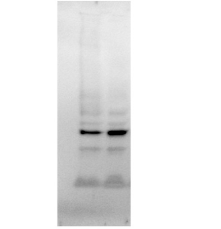
This image was generously provided by Loyda M. Meléndez at the University of Puerto Rico Medical Sciences Campus.\\nA total of 30ug of monocyte-derived macrophage lysates were reduced and denatured in 1X Laemli Buffer with beta-mercaptoethanol. Samples were loaded into a TGX 4-20% gel, at 150V for 1 hour. Proteins were transferred to a PVDF membrane and probed for proteins of interest. The membrane was re-probed with Rabbit Anti-GAPDH Polyclonal Antibody (bs-0459R) in 5% milk at 1:250 dilution overnight at 4°C. The next day, the membrane was washed with TBS-T and incubated with goat anti-rabbit HRP conjugated secondary antibody for 1 hour at room temperature. The membrane was exposed for 18 seconds.\\n
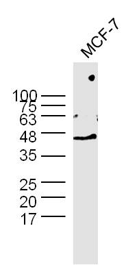
MCF-7 cell lysates probed with Rabbit Anti-GAPDH Polyclonal Antibody (bs-0459R) at 1:300 overnight at 4˚C. Followed by a conjugated secondary antibody (bs-0295G-HRP ) at 1:5000 for 90 min at 37˚C.
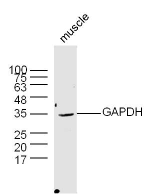
Mouse muscle lysates probed with Rabbit Anti-GAPDH Polyclonal Antibody (bs-0459R) at 1:300 overnight at 4˚C. Followed by a conjugated secondary antibody (bs-0295G-HRP ) at 1:5000 for 90 min at 37˚C.