Imprint® Monoclonal Anti-5-methylcytosine (33D3) antibody produced in mouse
| Name | Imprint® Monoclonal Anti-5-methylcytosine (33D3) antibody produced in mouse |
|---|---|
| Supplier | Sigma-Aldrich |
| Catalog | SAB4800001 |
| Prices | $544.00 |
| Sizes | 100 µg |
| Host | Mouse |
| Clonality | Monoclonal |
| Clone | 33D3 |
| Applications | MeDIP ICC/IF FC ELISA |
| Species Reactivities | Mouse, Human |
| Description | Mouse Monoclonal |
| Conjugate | unconjugated |
| Supplier Page | Shop |
Product images
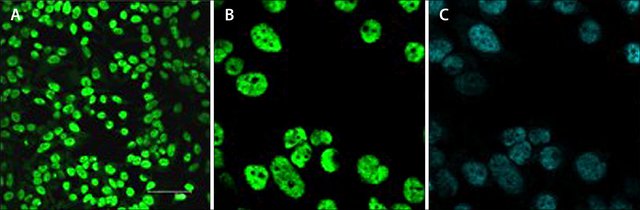 Immunofluorescence
Anti-5-methylcytosine: Cat. No. SAB4800001: HeLa cells were stained with 5-methylcytosine Mouse monoclonal antibody, 33D3 clone, and nuclei were counterstained with DAPI. Cells were methanol fixed for 10 minutes at -20 °C. Cells were re-hydrated in PBS and then treated with 2N HCl for 30 minutes at 37 °C. After two washes in borate buffer, cells were blocked with 1% BSA containing PBS.
Figure A: Cells were immunofluorescent labeled with the antibody diluted 1:100 in the blocking solution and incubated with the cells during one hour at room temperature in the dark, followed by a Goat Anti-Mouse FITC conjugated antibody. Scale bar is 75 μm.
Figure B: Enlarged picture corresponding to a region from the left panel, as indicated in Figure A.
Figure C: Nuclei were DAPI stained to specifically label the DNA. The region shown is the same as seen in Figure B.
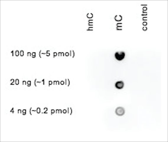 Immunoblotting
Anti-5-methylcytosine: Cat. No. SAB4800001: Dot blot analysis of the 5-methylcytosine monoclonal antibody with cytosine, C, methylcytosine, mC, and hydroxymethylcytosine, hmC, PCR controls. 100 to 4 ng, equivalent of 5 to 0.2 pmol of C-base, of hmC, mC and C PCR controls were spotted on a membrane. The membrane was incubated with 4 μg/mL of the antibody, followed by an HRP conjugated secondary antibody. The membrane was exposed for 30 seconds.
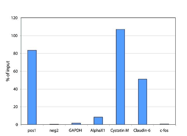 ChIP Results Chart
Anti-5-methylcytosine: Cat. No. SAB4800001: Methyl DNA IP assays were performed using DNA from MCF7 cells to analyze different genomic regions. The IP was performed including positive and negative controls, methylated and unmethylated DNA respectively, together with human DNA sample. IP minutesd material was analyzed by qPCR using primer pairs targeting specific DNA.
Columns, left to right,
1. methylated DNA positive control, pos1,
2. unmethylated DNA negative control, neg2,
3. GAPDH promoter, GAPDH,: Unmethylated region, no signal expected.
4. X-linked alpha-satellites, AlphaX1,: Methylated region, signal expected.
5. Cystatin M promoter, Cystatin M,: Methylated region in MCF7 cells, signal expected.
6. Claudin-6: Methylated region in MCF7 cells, signal expected.
7. c-fos: Unmethylated region, no signal expected.