CDIPT-Antibody-Center
| Name | CDIPT-Antibody-Center |
|---|---|
| Supplier | Abgent, a WuXi AppTec company |
| Catalog | AP9044c |
| Prices | $99.00, $295.00 |
| Sizes | 80 µl, 400 µl |
| Host | Rabbit |
| Clonality | Polyclonal |
| Isotype | Rabbit Ig |
| Applications | WB IHC-P FC ELISA |
| Species Reactivities | Human, Mouse |
| Antigen | 99-125 aa |
| Purity/Format | Purified polyclonal antibody supplied in PBS with 0.09% (W/V) sodium azide. This antibody is purified through a protein A column, followed by peptide affinity purification. |
| Description | Rabbit Polyclonal |
| Gene | CDIPT |
| Supplier Page | Shop |
Product images
 Western blot analysis of CDIPT Antibody (Center) (Cat. #AP9044c) in mouse stomach tissue lysates (35ug/lane). CDIPT (arrow) was detected using the purified Pab.
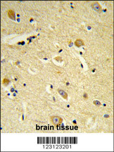 Formalin-fixed and paraffin-embedded human brain tissue reacted with CDIPT Antibody (Center), which was peroxidase-conjugated to the secondary antibody, followed by DAB staining. This data demonstrates the use of this antibody for immunohistochemistry; clinical relevance has not been evaluated.
 CDIPT Antibody (Center) (Cat. #AP9044c) flow cytometry analysis of MCF-7 cells (bottom histogram) compared to a negative control cell (top histogram).FITC-conjugated goat-anti-rabbit secondary antibodies were used for the analysis.