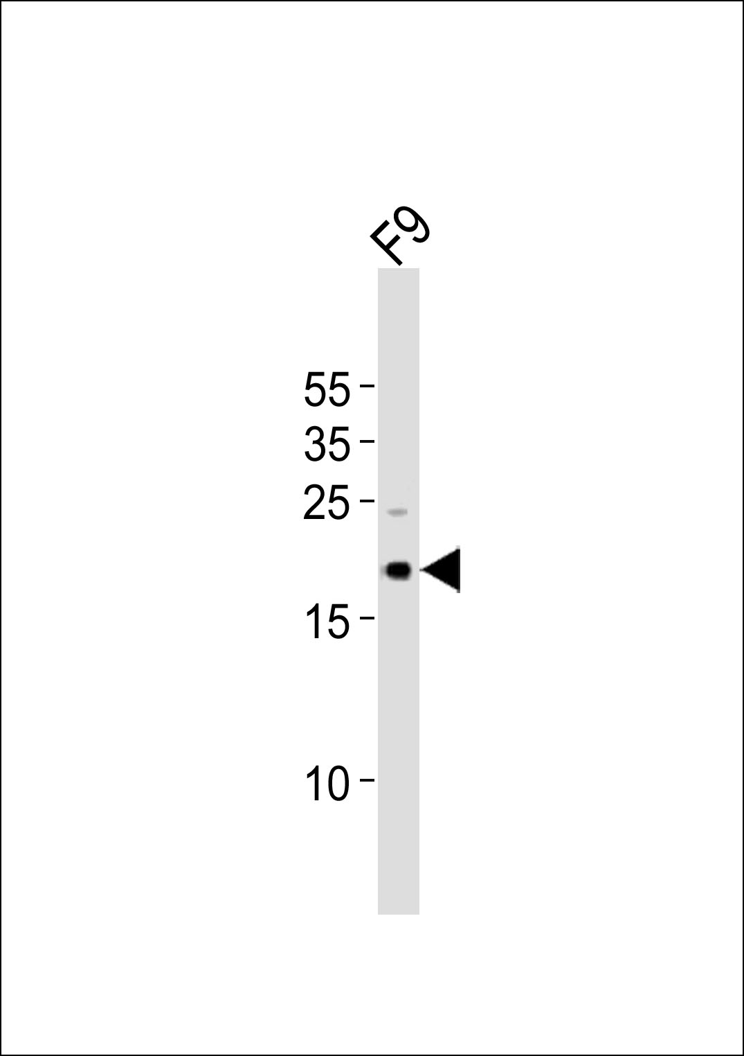
Western blot analysis of lysate from mouse F9 cell line, using (Mouse) Tdgf1 Antibody (N-term)(Cat. #AP21040a). AP21040a was diluted at 1:1000. A goat anti-rabbit IgG H&L(HRP) at 1:10000 dilution was used as the secondary antibody. Lysate at 20ug.
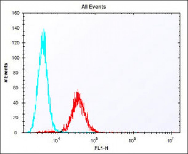
Overlay histogram showing F9 cells stained with AP21040a (red line). The cells were fixed with 2% paraformaldehyde (10 min) and then permeabilized with 90% methanol for 10 min. The cells were then icubated in 2% bovine serum albumin to block non-specific protein-protein interactions followed by the antibody (AP21040a, 1:25 dilution) for 60 min at 37ºC. The secondary antibody used was Alexa Fluor® 488 goat anti-rabbit lgG (H+L) (1583138) at 1/400 dilution for 40 min at 37ºC. Isotype control antibody (blue line) was rabbit IgG1 (1μg/1x10^6 cells) used under the same conditions. Acquisition of >10, 000 events was performed.