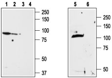
Western blot analysis of ND7/23 cell line membrane (1,3), RBL lysates (2,4) and rat brain membrane (5,6): 1,2,5. Anti-TRPV2 (extracellular) antibody (#AG1345), (1:200). 3,4,6. Anti-TRPV2 (extracellular) antibody, preincubated with the control peptide antigen.
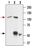
Immunoprecipitation of rat basophilic leukemia (RBL) cell lysate: 1. RBL lysate. 2. Lysate immunoprecipitated with Anti-TRPV2 (extracellular) antibody (#AG1345), (6 mg). 3. Lysate immunoprecipitated with pre-immune rabbit serum. The upper arrow indicates TRPV2 while the lower arrow indicates the IgG heavy chain. Western blot analysis was performed with Anti-TRPV2 (extracellular) antibody.

Expression of TRPV2 in mouse DRG Immunohistochemical staining of TRPV2 in mouse dorsal root ganglion (DRG) using Anti-TRPV2 (extracellular) antibody (#AG1345). A. TRPV2 (green) appears in patches along the perimeter of the DRG (arrows). B) Neurons containing neurofilament 200 (red) are scattered in the DRG, also in patches (arrows). C. A merge of the two panels shows that the spatial distribution of neurofilament 200 and TRPV2 expression overlaps. However, DRGs showing robust expression of neurofilament 200 do not contain TRPV2.
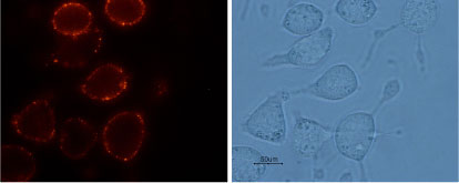
Expression of TRPV2 in RBL cells Immunocytochemical staining of intact living rat basophilic leukemia (RBL) cells with Anti-TRPV2 (extracellular) antibody (#AG1345), (1:100), followed by goat anti-rabbit-AlexaFluor-550 secondary antibody (red), (x100).

Indirect flow cytometry analysis of intact living RBL cells: Black Unstained cells.___ Cells + Anti-TRPV2 (extracellular) antibody (#AG1345).