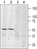
Western blot analysis of mouse brain (lanes 1 and 3) and rat brain (lanes 2 and 4) lysates: 1, 3. Anti-β3-Adrenoceptor (extracellular) antibody (#AG1379), (1:200). 2, 4. Anti-β3-Adrenoceptor (extracellular) antibody, preincubated with the control peptide antigen.
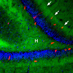
Expression of β3-Adrenoceptor in rat hippocampus Immunohistochemical staining of rat hippocampal dentate gyrus using Anti-β3-Adrenoceptor antibody (#AG1379), (1:100), (green). β3-Adrenoceptor is strongly expressed in the hilus (H) and in the outer molecular layer (arrows). The distribution overlaps the entire layers, but is not restricted to nerve cells (stained red with mouse anti Parvalbumin). DAPI is used as the counterstain (blue).