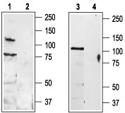
Western blot analysis of rat DRG (lanes 1 and 2) and rat brain (lanes 3 and 4) lysates: 1, 3. Anti-TRPV3 (extracellular) antibody (#AG1451), (1:200). 2, 4. Anti-TRPV3 (extracellular) antibody, preincubated with the control peptide antigen.
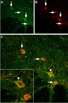
Expression of TRPV3 in mouse spinal cord Immunohistochemical staining of mouse spinal cord using Anti-TRPV3 (extracellular) antibody (#AG1451). A. TRPV3 (green) appears in neurons (vertical arrows) in the ventral horn of the mouse spinal cord. B. Motor neurons were stained with goat anti choline-acetyltransferase (red). In some motor neurons (A) TRPV3 was more intense (vertical arrow) whereas in others it was weak (horizontal arrows). C. Merged images of panels A and B. The inset in C magnifies one large motor neuron (vertical arrowhead). Note the punctate pattern of TRPV3 on the surface of the soma.
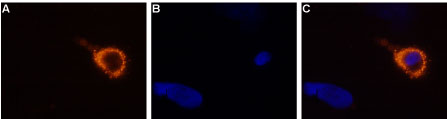
Expression of TRPV3 in rat DRG primary culture Immunocytochemical staining of living rat dorsal root ganglion (DRG) primary culture using Anti-TRPV3 (extracellular) antibody (#AG1451), (1:50-1:100), followed by goat anti-rabbit-AlexaFluor-555 secondary antibody (A). B. Nuclear staining with the cell-permeable dye Hoechst 33342. C. Merged image of panels A and B.