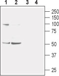
Western blot analysis of mouse (lanes 1 and 3) and rat (lanes 2 and 4) brain membranes: 1-2. Anti-Nicotinic Acetylcholine Receptor α2 (extracellular) antibody (#AG1422), (1:400). 3-4. Anti-Nicotinic Acetylcholine Receptor α2 (extracellular) antibody, preincubated with the control peptide antigen.
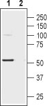
Western blot analysis of human SH-SY5Y neuroblastoma cell lysate: 1. Anti-Nicotinic Acetylcholine Receptor α2 (extracellular) antibody (#AG1422), (1:200). 2. Anti-Nicotinic Acetylcholine Receptor α2 (extracellular) antibody, preincubated with the control peptide antigen.
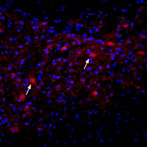
Expression of nAChRα2 in rat deep cerebellar nucleus Immunohistochemical staining of immersion-fixed, free floating rat brain frozen sections using Anti-Nicotinic Acetylcholine Receptor α2 (extracellular) antibody (#AG1422), (1:100). Staining reveals expression of nAChRα2 (red) in cells with neuronal outline (arrows point at some examples) in the red nucleus. DAPI is used as the counterstain (blue).
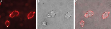
Expression of Nicotinic Acetylcholine Receptor α2 in rat PC12 pheochromocytoma cells Immunocytochemical staining of live intact rat PC12 pheochromocytoma cells. A. Extracellular staining of live cells with Anti-Nicotinic Acetylcholine Receptor α2 (extracellular) antibody (#AG1422), (1:50), followed by goat anti-rabbit-AlexaFluor-594 (red). B. Live image of the cells. C. Merge of the two images.