FOXO1-Antibody
| Name | FOXO1-Antibody |
|---|---|
| Supplier | Abgent, a WuXi AppTec company |
| Catalog | AO1677a |
| Prices | $385.00 |
| Sizes | 100 µl |
| Host | Mouse |
| Clonality | Monoclonal |
| Isotype | IgG1 |
| Clone | 3B6 |
| Applications | WB IHC ICC/IF ELISA |
| Species Reactivities | Human, Mouse |
| Antigen | Purified recombinant fragment of human FOXO1 expressed in E. Coli. |
| Description | Mouse Monoclonal |
| Gene | BPIFA2 |
| Supplier Page | Shop |
Product images
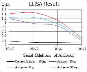 Black line: Control Antigen (100 ng); Purple line: Antigen(10ng); Blue line: Antigen (50 ng); Red line: Antigen (100 ng);
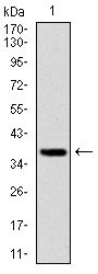 Figure 2: Western blot analysis using FOXO1 mAb against human FOXO1 (AA: 471-600) recombinant protein. (Expected MW is 39.3 kDa)
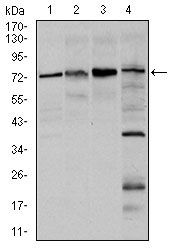 Figure 3: Western blot analysis using FOXO1 mouse mAb against Hela (1), HEK293 (2), MCF-7(3), and C6 (4) cell lysate.
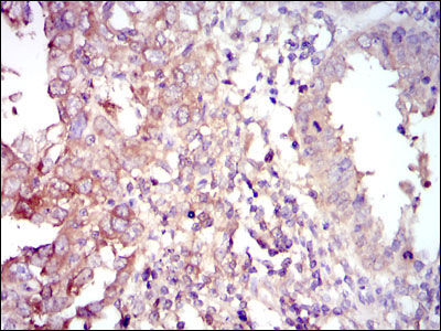 Figure 4: Immunohistochemical analysis of paraffin-embedded intima cancer tissues using FOXO1 mouse mAb with DAB staining.
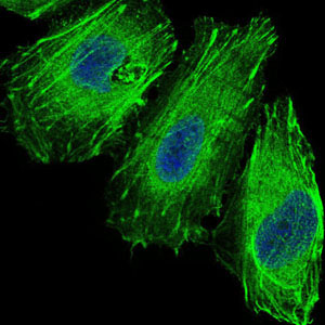 Figure 5: Immunofluorescence analysis of Hela cells using FOXO1 mouse mAb (green). Blue: DRAQ5 fluorescent DNA dye.