Anti-Apc7 antibody
| Name | Anti-Apc7 antibody |
|---|---|
| Supplier | Abcam |
| Catalog | ab4171 |
| Prices | $370.00 |
| Sizes | 100 µl |
| Host | Rabbit |
| Clonality | Polyclonal |
| Isotype | IgG |
| Applications | WB ICC/IF ICC/IF |
| Species Reactivities | Mouse, Human |
| Antigen | Synthetic peptide conjugated to KLH derived from within residues 100 - 200 of Human Apc7 |
| Blocking Peptide | Apc7 peptide |
| Description | Rabbit Polyclonal |
| Gene | ANAPC7 |
| Conjugate | Unconjugated |
| Supplier Page | Shop |
Product images
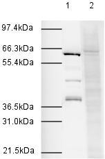 All lanes : Anti-Apc7 antibody (ab4171)Lane 1 : HeLa Nuclear ExtractLane 2 : HeLa Nuclear Extract with Apc7 peptide (ab13734) at 1 µg/mlLysates/proteins at 20 µg per lane.SecondaryLane 1 : Goat Anti-Rabbit IgG H&L (HRP) (ab6721) at 1/2000 dilutionLane 2 : Goat polyclonal to Rabbit IgG - H&L (HRP) (ab6721) at 1/2000 dilution
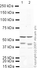 All lanes : Anti-Apc7 antibody (ab4171) at 1 µg/mlLane 1 : Heart (Mouse) Tissue LysateLane 2 : Spinal Cord (Mouse) Tissue LysateLysates/proteins at 10 µg per lane.SecondaryIRDye 680 Conjugated Goat Anti-Rabbit IgG (H+L) at 1/10000 dilutionPerformed under reducing conditions.
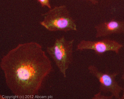 ICC/IF image of ab4171 stained HeLa cells. The cells were 4% formaldehyde fixed (10 min) and then incubated in 1%BSA / 10% normal goat serum / 0.3M glycine in 0.1% PBS-Tween for 1h to permeabilise the cells and block non-specific protein-protein interactions. The cells were then incubated with the antibody (ab4171, 10µg/ml) overnight at +4°C. The secondary antibody (green) was ab96899, DyLight® 488 goat anti-rabbit IgG (H+L) used at a 1/250 dilution for 1h. Alexa Fluor® 594 WGA was used to label plasma membranes (red) at a 1/200 dilution for 1h. DAPI was used to stain the cell nuclei (blue) at a concentration of 1.43µM.
Product References
How APC/C-Cdc20 changes its substrate specificity in mitosis. - How APC/C-Cdc20 changes its substrate specificity in mitosis.
Izawa D, Pines J. Nat Cell Biol. 2011 Mar;13(3):223-33.
The APC/C maintains the spindle assembly checkpoint by targeting Cdc20 for - The APC/C maintains the spindle assembly checkpoint by targeting Cdc20 for
Nilsson J, Yekezare M, Minshull J, Pines J. Nat Cell Biol. 2008 Dec;10(12):1411-20.