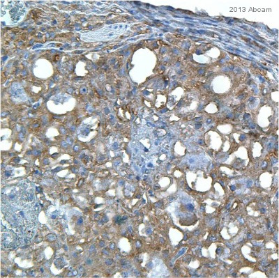
ab108251 staining Apg3 in Mouse uterus tissue sections by Immunohistochemistry (IHC-Fr - frozen sections). Tissue was fixed with formaldehyde and blocked with 5% BSA for 1 hour at 25°C. Samples were incubated with primary antibody (1/200 in 1% BSA in PBS) for 12 hours at 4°C. An undiluted HRP-conjugated Goat anti-rabbit IgG polyclonal was used as the secondary antibody.See Abreview
![All lanes : Anti-Apg3 antibody [EPR4801] (ab108251) at 1/10000 dilutionLane 1 : K562 cell lysateLane 2 : HL-60 cell lysateLane 3 : HeLa cell lysateLane 4 : Jurkat cell lysateLysates/proteins at 10 µg per lane.SecondaryHRP labelled goat anti-rabbit at 2000](http://www.bioprodhub.com/system/product_images/ab_products/2/sub_1/8154_Apg3-Primary-antibodies-ab108251-2.jpg)
All lanes : Anti-Apg3 antibody [EPR4801] (ab108251) at 1/10000 dilutionLane 1 : K562 cell lysateLane 2 : HL-60 cell lysateLane 3 : HeLa cell lysateLane 4 : Jurkat cell lysateLysates/proteins at 10 µg per lane.SecondaryHRP labelled goat anti-rabbit at 2000
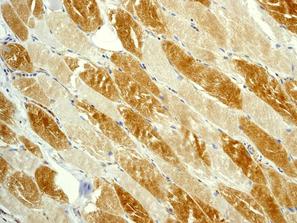
ab108251, at a 1/100 dilution, staining Human Apg3 in muscle, using Immunohistochemistry, Formalin/PFA-fixed paraffin-embedded tissue.

Immunofluorescent staining of HeLa cells using ab108251 at 1/100.