![All lanes : Anti-BRN3A antibody [EP1972Y] (ab81213)Lane 1 : LNCaPLane 2 : SW480Lane 3 : PC-3Lane 4 : Human brain lysate](http://www.bioprodhub.com/system/product_images/ab_products/2/sub_1/15700_BRN3A-Primary-antibodies-ab81213-10.jpg)
All lanes : Anti-BRN3A antibody [EP1972Y] (ab81213)Lane 1 : LNCaPLane 2 : SW480Lane 3 : PC-3Lane 4 : Human brain lysate
Immunohistochemistry analysis of paraffin-embedded Human placenta tissue using 1/100 ab81213.
Immunofluorescent staining of HeLa cells using 1/100 ab81213.
![All lanes : Anti-BRN3A antibody [EP1972Y] (ab81213) at 1/2000 dilutionLane 1 : Adult mouse cerebellum lysateLane 2 : Adult mouse lower brainstem lysateLysates/proteins at 20 µg per lane.SecondaryHRP-conjugated Mouse anti-Rabbit IgG monoclonal. at 1/10000 dilutionPerformed under reducing conditions.](http://www.bioprodhub.com/system/product_images/ab_products/2/sub_1/15703_BRN3A-Primary-antibodies-ab81213-15.jpg)
All lanes : Anti-BRN3A antibody [EP1972Y] (ab81213) at 1/2000 dilutionLane 1 : Adult mouse cerebellum lysateLane 2 : Adult mouse lower brainstem lysateLysates/proteins at 20 µg per lane.SecondaryHRP-conjugated Mouse anti-Rabbit IgG monoclonal. at 1/10000 dilutionPerformed under reducing conditions.
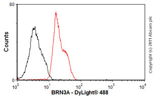
Overlay histogram showing SH-SY5Y cells stained with ab81213 (red line). The cells were fixed with 80% methanol (5 min) and then permeabilized with 0.1% PBS-Tween for 20 min. The cells were then incubated in 1x PBS / 10% normal goat serum / 0.3M glycine to block non-specific protein-protein interactions followed by the antibody (ab81213, 1/50 dilution) for 30 min at 22ºC. The secondary antibody used was DyLight® 488 goat anti-rabbit IgG (H+L) (ab96899) at 1/500 dilution for 30 min at 22ºC. Isotype control antibody (black line) was rabbit IgG (monclonal) (1µg/1x106 cells) used under the same conditions. Acquisition of >5,000 events was performed.