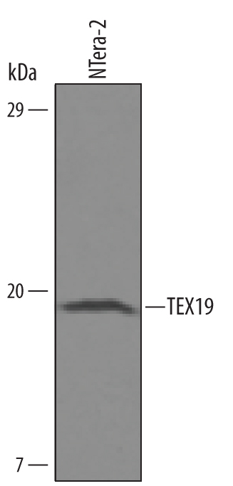
Detection of Human TEX19 by Western Blot. Western blot shows lysates of NTera-2 human testicular embryonic carcinoma cell line. PVDF Membrane was probed with 0.5 µg/mL of Human TEX19 Antigen Affinity-purified Polyclonal Antibody (Catalog # AF6319) followed by HRP-conjugated Anti-Sheep IgG Secondary Antibody (Catalog # HAF016 ). A specific band was detected for TEX19 at approximately 18 kDa (as indicated). This experiment was conducted under reducing conditions and using Immunoblot Buffer Group 8 .
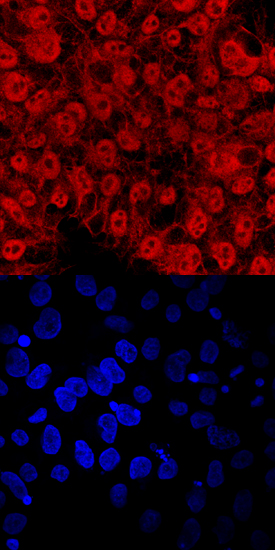
TEX19 in BG01V Human Stem Cells. TEX19 was detected in immersion fixed BG01V human embryonic stem cells using Human TEX19 Antigen Affinity-purified Polyclonal Antibody (Catalog # AF6319) at 10 µg/mL for 3 hours at room temperature. Cells were stained using the NorthernLights™ 557-conjugated Anti-Sheep IgG Secondary Antibody (red, upper panel; Catalog # NL010 ) and counterstained with DAPI (blue, lower panel). Specific staining was localized to nuclei and cytoplasm. View our protocol for Fluorescent ICC Staining of Cells on Coverslips .